Ventricular ejection and arterial elastic behaviour shape pulsatile pressure.
NORMAL PHYSIOLOGY / A CONNECTED LIVING CIRCUIT
Blood Pressure & Flow
Understand the push, the delivery and the feedback—without confusing one for another.
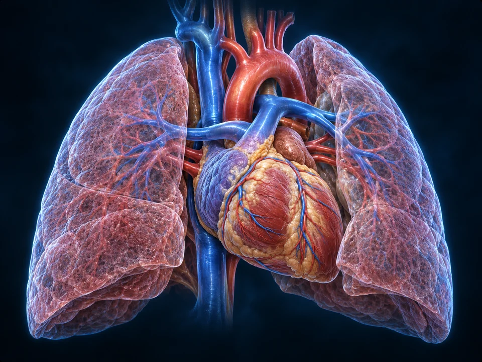
Start with the pump and the returning circulation. Look inside a resistance vessel, then follow how arterial stretch becomes a coordinated response.
30-SECOND OVERVIEW / PUMP → ROUTES → RETURN
The pump, the vessel walls and the returning blood work together.
Regional context · pumps and return
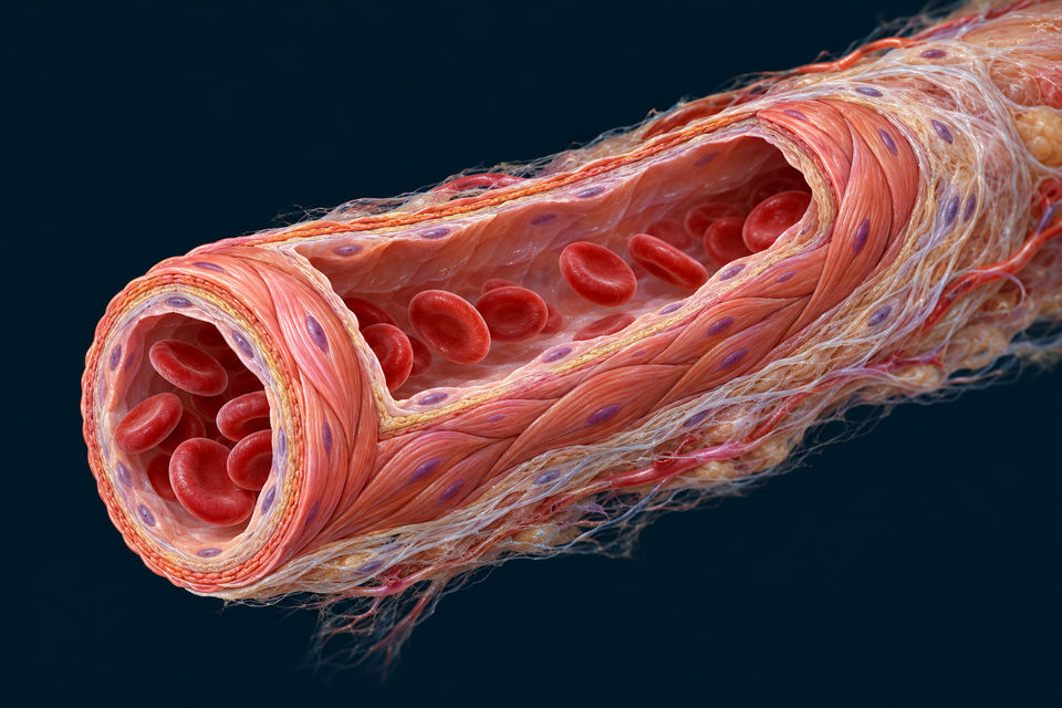
Different scale · the adjustable wall
Structure notes · further context
- Open blood lumen
- Volume flow concerns blood moving through this inside space.
- Endothelial lining
- The blood-facing layer separates the lumen from the surrounding muscular wall.
- Circumferential muscle
- Smooth-muscle activity changes calibre. The muscular cells lie outside the endothelial boundary.
- Outer connective tissue
- External collagenous support belongs outside the muscle; it is not another blood-filled lumen.
Resistance and local activity influence how flow reaches tissue beds.
Return, neural feedback and renal-volume regulation connect the whole system.
Sources for this relationship: OpenStax · Flow, pressure and resistance · OpenStax · Vascular homeostasis
01 / THREE DIFFERENT QUESTIONS
Pressure pushes. Flow delivers. Speed describes motion.
Pressure is force per area. Volume flow is how much blood crosses a section per time. Mean speed depends on that flow and the cross-sectional area being considered. They are related quantities, not three names for the same thing.
Force per area at a named location
Specify the compartment and reference. An arterial value is not a capillary or venous value. A pressure difference can help drive flow.
An arterial pulse is a propagating pressure disturbance, not a parcel of blood racing from the heart to the fingertip with each beat. Do not infer red-cell travel from the speed at which a pulse is detected.
Many capillaries in parallel create a large combined cross-sectional area. A single capillary’s narrow diameter alone does not establish the speed or resistance of the whole exchange network.
Sources for this relationship: OpenStax · Flow, pressure and resistance · CV Physiology · Aortic and arterial pulse pressure
02 / A PULSE IS NOT ITS AVERAGE
Systolic, diastolic and mean pressure describe different features.
Systolic pressure is the cycle’s high arterial value; diastolic pressure is its low value. Pulse pressure is their difference. Mean arterial pressure is a time average over the cycle, not simply the midpoint of the two extrema.
Ejection and arterial elastic behaviour shape the pulse. Recoil helps sustain arterial flow between ejections. The arterial pressure remaining during diastole does not mean the ventricle is still ejecting.
Timing matters when estimating a cycle average. This lesson provides no diagnostic thresholds, individual target or interpretation of a home reading. The existing Heart course shows valve and ventricular phases separately.
Sources for this relationship: Physiology, Mean Arterial Pressure · CV Physiology · Aortic and arterial pulse pressure
03 / NAME THE UPSTREAM AND DOWNSTREAM SIDES
Flow depends on a pressure difference, not one isolated number.
For a chosen vascular pathway in an averaged description, Q = ΔP/R. For the systemic circuit, the relevant overall difference is mean arterial pressure minus right-atrial pressure. The resistance must refer to that same chosen pathway.
Rearranging gives MAP − right-atrial pressure = cardiac output × systemic vascular resistance in the simplified steady description. Omitting the downstream term is an approximation, not a universal identity.
A local flow can fall at the same upstream pressure if local resistance increases. Conversely, maintaining a flow through greater resistance requires a larger pressure difference. Real cardiac and vascular responses are coupled, so the body does not independently fix every variable as in a classroom comparison.
Sources for this relationship: Physiology, Mean Arterial Pressure · Control of Cardiac Output · Venous return · Physiology, Peripheral Vascular Resistance
04 / A LIVING WALL CHANGES THE ROUTE
Smooth muscle surrounds the blood-facing lining.
Small muscular resistance vessels change calibre through vascular smooth-muscle activity. Locate the lumen, thin endothelial lining, surrounding muscle and external connective tissue before using a mathematical tube comparison.
Structure notes · further context
- Open blood lumen
- Volume flow concerns blood moving through this inside space.
- Endothelial lining
- The blood-facing layer separates the lumen from the surrounding muscular wall.
- Circumferential muscle
- Smooth-muscle activity changes calibre. The muscular cells lie outside the endothelial boundary.
- Outer connective tissue
- External collagenous support belongs outside the muscle; it is not another blood-filled lumen.
Change one ideal assumption at a time.
Hold tube length, viscosity and driving pressure difference fixed. Compare three chosen radii with the reference radius of 1.0. These are relative inputs, not measurements or preferred vessel sizes.
Relative resistance: 1.00
R/R₀ = 1 / 1.0⁴
Relative flow: 1.00
Q/Q₀ = 1.0⁴ at fixed ΔP
Branching, compliance, red-cell behaviour and feedback are outside this comparison. It cannot predict a personal pressure response.
The new preparation is an enlarged teaching cutaway, not microscopy. Its cells and layers explain a structural relationship; the pixels do not measure a person’s vessel diameter or identify a molecular channel.
Under the ideal assumptions of steady laminar flow in a rigid circular tube with fixed length and viscosity, resistance varies inversely with radius to the fourth power. Real blood, compliant branching vessels and changing muscle activity require more than this equation.
Sources for this relationship: Physiology, Peripheral Vascular Resistance · Histology Guide · Arterioles and venules, MH024–025
05 / STORAGE IS NOT RESISTANCE
A compliant wall accommodates volume; a resistance opposes flow.
Compliance is the change in contained volume per change in pressure. Elastic arteries accommodate pulsatile input and recoil; the venous compartment can store a substantial changing volume at lower pressure.

Structure notes · further context
- Arterial lumen
- Broad named relationship checked against independent normal anatomy sources and the visible new illustration. Fine surface/granule/layer dimensions and hidden detail are illustrative / not independently verified. Human medical review: not performed.
- Intimal context
- Broad named relationship checked against independent normal anatomy sources and the visible new illustration. Fine surface/granule/layer dimensions and hidden detail are illustrative / not independently verified. Human medical review: not performed.
- Muscular media
- Broad named relationship checked against independent normal anatomy sources and the visible new illustration. Fine surface/granule/layer dimensions and hidden detail are illustrative / not independently verified. Human medical review: not performed.
- Outer connective tissue
- Broad named relationship checked against independent normal anatomy sources and the visible new illustration. Fine surface/granule/layer dimensions and hidden detail are illustrative / not independently verified. Human medical review: not performed.
- Venous wall
- Broad named relationship checked against independent normal anatomy sources and the visible new illustration. Fine surface/granule/layer dimensions and hidden detail are illustrative / not independently verified. Human medical review: not performed.
- Venous lumen
- Broad named relationship checked against independent normal anatomy sources and the visible new illustration. Fine surface/granule/layer dimensions and hidden detail are illustrative / not independently verified. Human medical review: not performed.
The retained image compares a muscular artery with a vein. It is not a before-and-after constriction pair and is not a substitute for the specialised elastic structure of the aorta.
Resistance and compliance can change together in living vessels, but they answer different questions. “A wider-looking vessel” alone does not reveal its pressure, flow or compliance from a still illustration.
Sources for this relationship: CV Physiology · Vascular compliance · CV Physiology · Aortic and arterial pulse pressure
06 / A WALL CAN REPORT ITS STRETCH
The carotid sinus is a vessel region, not the carotid body.
The carotid sinus is commonly a widened region near the start of the internal carotid artery. Stretch-sensitive nerve endings lie in its wall context. The nearby carotid body is a separate chemosensory structure outside the artery.

Structure notes · further context
- Common carotid
- The common arterial trunk divides towards internal and external routes.
- External carotid route
- This cervical route gives branches in the neck; the visible short branches are not a complete named inventory.
- Carotid sinus region
- A widened region near the proximal internal carotid provides arterial wall-stretch sensing context.
- Separate carotid body
- The small external chemosensory structure is different from the stretch-sensitive arterial sinus.
- Wall sensory endings
- Terminals in outer-wall context respond to deformation. They do not float in the blood lumen.
- Sensory route
- Carotid-sinus input proceeds through a branch of cranial nerve IX towards medullary integration, beyond this local view.
Gold arrow: afferent information towards the proximal sensory route, not blood flow or a measured nerve impulse.
Tonic sensory input participates in a balanced system.
The system continuously samples arterial stretch. Current autonomic output also reflects other inputs; baseline does not mean receptors are silent.
- Arterial wall stretch
- Carotid IX / aortic X afferents
- Medullary integration
- Coordinated heart and vascular output
The local carotid plate shows one input site. Aortic-arch input is located on the retained overview; its detailed terminals and the medullary nuclei are not exposed by this view.
The illustration opens a small wall window for visibility. Sensory terminals belong to the wall, not the blood lumen. Their generated pattern is not a complete terminal-branch map or measured histological field.
Carotid-sinus afferents travel through a branch of cranial nerve IX. Aortic-arch afferents travel through cranial nerve X. Both contribute to medullary integration; the two sensory routes must not be swapped or described as motor commands entering an artery.
Sources for this relationship: Anatomy, Head and Neck: Carotid Sinus · Physiology, Baroreceptors · NCBI Bookshelf
07 / INPUT → INTEGRATION → COORDINATED OUTPUT
Compare the direction of an isolated reflex response.
Arterial wall stretch changes sensory input. Medullary circuits coordinate cardiac vagal and sympathetic output to the heart and blood vessels. This rapid feedback buffers disturbances; it is not a complete prediction of a person’s blood pressure.
The selectable cases isolate the arterial baroreflex. Ongoing breathing, movement, local demands and hormonal signals also affect the resulting response. There is no claim that every organ constricts or that a chosen nerve firing rate gives a particular measured pressure.
Sustained changes can shift the operating relationship of baroreceptors. Short-term reflex buffering should not be presented as the sole controller of long-term blood volume or pressure.
Return to the three reflex comparisons and local sensor atlas →
Sources for this relationship: Physiology, Baroreceptors · NCBI Bookshelf · OpenStax · Vascular homeostasis
08 / THE TISSUE HAS ITS OWN INPUTS
A local supply response and a whole-body pressure reflex are different.
Resistance vessels also respond to local conditions. Metabolic signals can help match tissue activity with delivery; a myogenic response changes smooth-muscle activity following changes in wall stretch.
Endothelial cells can respond to shear and chemical signals, communicating with surrounding smooth muscle. Nitric oxide is one participant, not a universal single switch for every organ.
These mechanisms interact with neural and circulating signals. Their relative contributions differ between tissues and conditions. The cardiovascular system is not one common tap delivering the same fraction to every organ at all times.
Sources for this relationship: Skeletal Muscle Circulation · Local vascular tone · Regulation of Tissue Oxygenation · Local flow control
09 / DELIVERY MUST BE FOLLOWED BY RETURN
The pump and the return pathway work as one circulation.
Venous volume and vascular tone affect how blood is distributed between stored and returning compartments. Right-atrial pressure is a downstream boundary for systemic return and also a filling relationship for the heart.
Muscle activity and respiratory pressure changes can assist venous return in suitable anatomical settings. Valves in many limb veins help limit backward flow; they are passive leaflets, not extra contracting hearts.
Mean output and return must match in a sustained steady state. A short-lived change can redistribute blood between compartments, so one selected pump number is not an independent permanent flow imposed on the whole circulation.
Sources for this relationship: Control of Cardiac Output · Venous return · CV Physiology · Vascular compliance
10 / RAPID BUFFERING MEETS VOLUME REGULATION
Kidneys, hormones and blood vessels contribute at different scales.
Renal sodium and water handling help determine extracellular volume, while neural and hormonal pathways affect vascular and renal behaviour. A quick baroreflex response does not replace these longer-term relationships.
Angiotensin and aldosterone have different targets and actions. Aldosterone affects epithelial transport; it is not a pressure wave travelling down an artery. Existing kidney and adrenal courses explain those mechanisms in detail.
Timing is not a strict two-box division: mechanisms overlap and influence each other. This chapter links the mature courses rather than duplicating their complete images or offering advice about salt, fluids or medication.
Sources for this relationship: OpenStax · Vascular homeostasis · Physiology, Mean Arterial Pressure
GO DEEPER / TISSUE DEFORMATION BECOMES A SIGNAL
A sensory ending converts mechanical change into neural activity.
Baroreception is mechanosensation. Deformation affects excitability in sensory endings; the nervous system carries information rather than transporting the sensed blood pressure as a material.
In a 2018 mouse study, removing both Piezo1 and Piezo2 from specified sensory neurons abolished the tested drug-induced reflex and aortic nerve activity. Optogenetic activation supplied a separate functional test.
That experimental evidence supports a mechanosensing role. It does not certify a generated human channel structure, establish the exact contribution of every human sensory subtype or supply a treatment instruction. The atlas shows regional anatomy, not proteins at molecular resolution.
Sources for this relationship: Zeng et al. · PIEZO mechanosensing and the baroreflex (2018) · Physiology, Baroreceptors · NCBI Bookshelf
REAL OBSERVATION / KEEP EVIDENCE TYPES DISTINCT
Compare acquired tissue with the generated teaching preparation.
The institutional MH024–025 mesentery guide offers acquired arteriole and venule sections and stain comparisons. Locate the endothelial boundary and surrounding muscle before comparing the enlarged atlas.
The page’s slide identity and preparation belong to that source. Species is not certified here. No external slide pixels are embedded, and no generated image is called a photomicrograph.
A tissue section can reveal wall organisation; it does not by itself measure living pressure or flow. A pressure waveform records a different variable, and a cuff estimate is a measurement method rather than direct visualisation of vessel-wall forces.
Sources for this relationship: Histology Guide · Arterioles and venules, MH024–025 · OpenStax · Flow, pressure and resistance
Check the variable, the boundary and the evidence.
If a pulse travels quickly, must the same red cell travel that distance at that speed?
No. A pressure disturbance and bulk blood transport are different phenomena.
What is missing from “higher resistance means higher pressure”?
Which pressure, which pathway and what is held fixed? At fixed flow, a greater resistance requires a larger pressure difference. At fixed pressure difference, it lowers flow.
Are the two vessels in the retained wall plate relaxed and constricted states?
No. They are different vessel types: a representative muscular artery and vein.
Does the carotid body shown near the fork measure arterial stretch?
The stretch-sensitive carotid sinus is a vessel region. The separate carotid body is primarily chemosensory.
Which cranial nerves carry the two major arterial sensory routes?
Carotid sinus: IX. Aortic arch: X. These are afferent routes to medullary integration, not the motor efferent limbs.
Can the radius comparison predict a personal pressure response?
No. It isolates an ideal rigid tube while fixing other assumptions. A branching compliant circulation with feedback is not that model.
Sources and teaching scope
Normal physiology reviewed against the sources below on 21 October 2026. Existing book inventory guided vascular organisation, linked pumping and pressure literacy. The text and illustrations are original or retained qualified project work; no reference-book prose, artwork or external clinical pixels are copied.
- OpenStax · Flow, pressure and resistanceNormal quantities and vascular organisation; no diagnostic ranges or clinical instructions reproduced.
- OpenStax · Vascular homeostasisNeural, endocrine and local coordination. Disease/treatment material excluded.
- Physiology, Baroreceptors · NCBI BookshelfStretch-sensitive afferents, IX/X distinction, medullary integration and reflex adaptation.
- Anatomy, Head and Neck: Carotid SinusBifurcation and proximal internal-carotid sinus anatomy only. Its combined-IX statement about arch afferents is not adopted.
- Physiology, Mean Arterial PressureCycle-averaged arterial pressure and contributions from output and resistance, not a personal target.
- Physiology, Peripheral Vascular ResistanceResistance-vessel role and ideal-tube radius dependence; definition clarified as pressure difference divided by volume flow.
- CV Physiology · Vascular complianceVolume change per pressure change and arterial versus venous storage.
- CV Physiology · Aortic and arterial pulse pressurePulsatile ejection, arterial elastic behaviour and pulse pressure.
- Skeletal Muscle Circulation · Local vascular toneMyogenic, metabolic, endothelial and conducted mechanisms, with tissue-specific scope.
- Regulation of Tissue Oxygenation · Local flow controlLocal matching of supply and tissue demand, not a universal fixed flow percentage.
- Control of Cardiac Output · Venous returnFilling pressure, venous volume/tone and return as part of a coupled circulation; numerical examples not reproduced.
- Zeng et al. · PIEZO mechanosensing and the baroreflex (2018)Mouse genetic and optogenetic experiments; does not certify a human molecular atlas or treatment.
- Histology Guide · Arterioles and venules, MH024–025Externally linked acquired tissue observation and stain comparison; no slide pixels copied and no assumed species.
This educational chapter does not interpret a personal reading, set a treatment target or recommend medication. AI-assisted source and anatomy checks are not physician review. Generated microscopic detail is not acquired tissue, and the ideal-tube arithmetic is not a cardiovascular patient model.