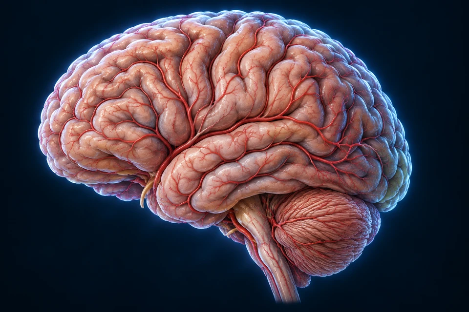
When brain blood flow is blocked—or a vessel bleeds.
Stroke injures brain tissue through loss of blood flow or bleeding. Fast recognition matters because affected networks may stop supporting movement, language, vision or other functions.
Sudden loss of balance or coordination; new trouble walking.
E Eyes
Sudden loss or change of vision, including double vision.
F Face
Sudden facial weakness, numbness or an uneven smile.
A Arms
Sudden weakness or numbness, often on one side; an arm may drift.
S Speech
Sudden slurring, trouble speaking or difficulty understanding.
T Time
Call local emergency services immediately. Note onset or last known well.
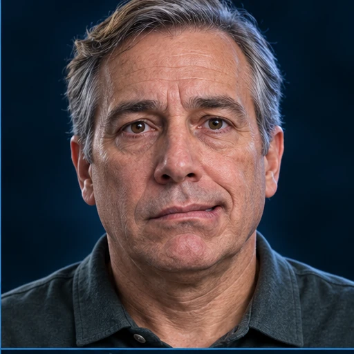
Original staged illustrations of recognition and emergency action, not patient photographs. A still image cannot establish a sudden symptom: ask about new changes. Eye appearance alone does not prove vision loss; facial appearance alone does not diagnose stroke.
Other sudden warning signs
Leg weakness or numbness, confusion, and a severe unexplained headache can also occur. This memory aid is not a diagnostic test or a complete checklist. A person can have stroke without the classic face, arm and speech pattern.
Symptoms stopped? Still an emergency.
A brief episode may be a transient ischemic attack (TIA), stroke or another condition. Recovery of symptoms does not establish the diagnosis or make it safe to wait. Emergency assessment can identify the cause and reduce future risk.
Call an ambulance through your local emergency service. Do not drive yourself. Note when symptoms began, or when the person was last known to be well. Do not take aspirin on your own for suspected stroke: it may worsen bleeding.
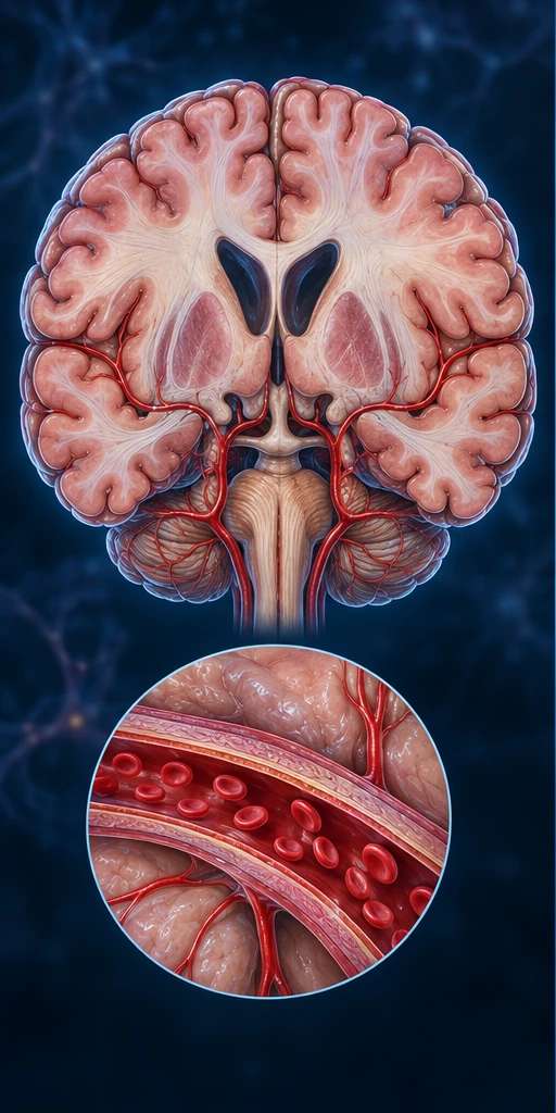
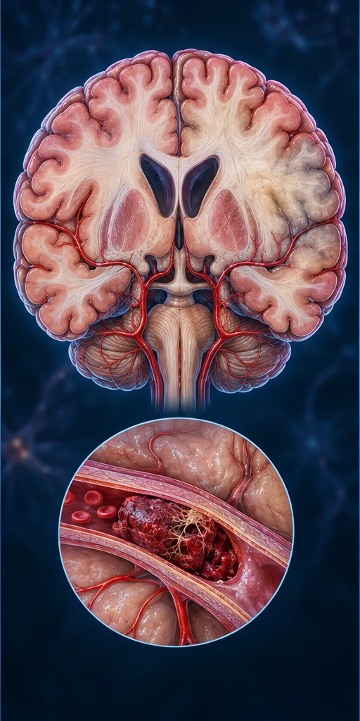
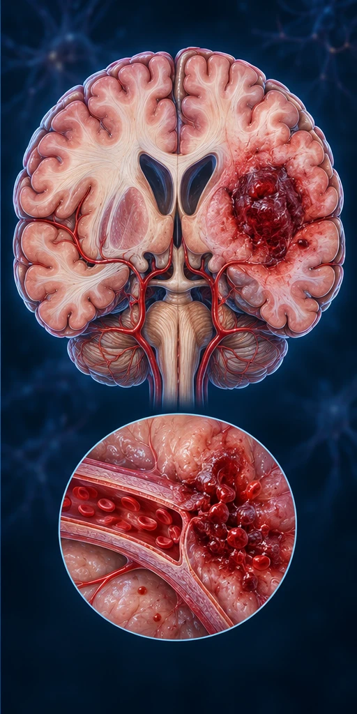
Brain cells need oxygen and nutrients carried by blood. An intact vessel keeps blood moving within its lumen. Loss of delivery and blood outside the vessel injure tissue through different mechanisms.
Normal delivery
Blood stays within a patent vessel. Oxygen and glucose reach tissue through the microcirculation.
Ischemic stroke
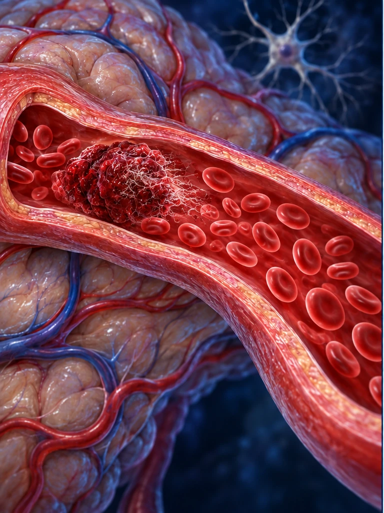
The clot is inside the vessel lumen. Reduced downstream delivery can injure connected brain tissue.
Intracerebral hemorrhage
Blood is outside the vessel, within tissue. A collection, pressure and blood products can add injury.
Stylized cerebral cutaway comparison with vessel enlargements. Hypothetical tissue changes; not a patient scan, exact anatomical section or arterial-territory map. The pale region is not a measured penumbra. Frozen blood cells do not show actual flow. The bleeding example is ICH, not SAH.
Ischemia: delivery falls
Thrombus means a clot forming at the site; embolus means material travelling to block another vessel. Small penetrating vessels can also become diseased. Reduced supply may disrupt a particular region, rather than the entire brain equally.
Hemorrhage: location matters
Intracerebral hemorrhage (ICH) is bleeding within brain tissue. Subarachnoid hemorrhage (SAH) is bleeding into the fluid-containing space around the brain; a ruptured aneurysm is one cause. These are not interchangeable labels or treatment pathways.
Location, extent and connected networks shape the effects.
The same symptom can arise from different lesions. A pictured lesion cannot predict an individual’s symptoms or determine the stroke type.
Coronal teaching example · viewer side is not a patient-side label. Exact nuclei, vessel of origin, lesion volume and tissue displacement are not independently resolved in this generated image. It is not a clinical CT/MRI or a universal symptom map.
Different networks, different changes
Motor and sensory pathways may cause weakness or altered sensation, often on the opposite body side. Language, visual, attention and coordination networks may also be affected. Brainstem injury can disturb several linked functions. The pattern requires clinical assessment.
In ischemia, some tissue becomes irreversibly injured while other under-supplied tissue may remain salvageable. Collateral flow and time influence this balance; it is not a fixed ring or universal countdown. Specialist assessment and imaging help guide treatment.
Bleeding can evolve
A hematoma may expand. Swelling, pressure and blood products add injury; blood or compression can sometimes disrupt fluid circulation. Repeated assessment matters even after the first scan.
From circulation to cellular energy
Why disrupted delivery threatens a neuron
Oxygen and glucose support ATP production. Loss of energy can disrupt ion regulation and promote swelling. The tissue view and two enlargements locate the supply, energy and membrane interfaces. They show interacting mechanisms, not a fixed sequence of cell death.
Several interfaces interact; this is not a timetable.
Gold outlines locate selected existing landmarks; they do not measure blood flow, ATP or ion movement. All four anatomical names remain visible. The other cellular relationships continue to matter in every selection.
Supply and cellular energy
Less oxygen and glucose limits oxidative energy production. ATP-dependent ion pumps can no longer sustain normal gradients. The capillary, mitochondrion and membrane are different observational scales, not consecutive stages.
Ion and transmitter imbalance
Depolarization and impaired glutamate handling can drive excessive excitatory signalling and calcium entry. Sodium, other ions and water shifts contribute to cell swelling; calcium can activate damaging enzymes.
Interacting injury
Mitochondrial dysfunction, oxidative stress, inflammation and blood–brain barrier changes can reinforce injury. Neurons, glia and vascular cells respond differently. Flow, severity, duration and reperfusion influence the outcome.
Generated neurovascular teaching context with separate scale enlargements, not histology or a measured injured specimen. Cell branching, endothelial contacts, organelle sizes and protein forms are illustrative. No specific channel or pump conformation, exact glial identity, oxygen value or irreversible damage is inferred from the shapes. Astrocytes normally support the blood–brain interface; this artwork does not resolve their complete endfoot coverage.
Mechanism summary: Reduced delivery can lower usable energy and disturb ion homeostasis, contributing to stress and injury. These are interacting relationships, not a universal timetable for every cell.
Compare the retained whole-cell stress illustration
Conceptual cell-stress comparison, not histology. Both panels contain blood cells; reduced delivery is explained in the text, not inferred from their count. Organelles, branching and swelling proportions are illustrative.
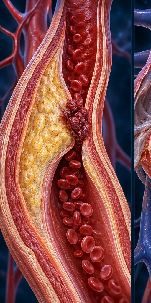
Atherosclerotic plaque can narrow an artery or promote a clot. Long-term small-vessel injury is another pathway. These explain an event’s mechanism; a risk factor describes increased likelihood.
Heart-to-brain connection
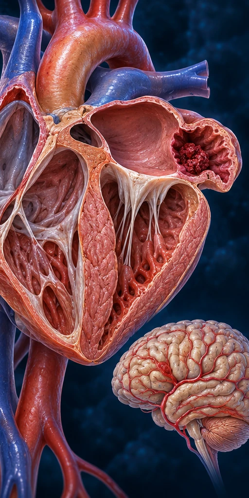
In atrial fibrillation, disturbed atrial contraction can allow a clot to form and travel to cerebral circulation. AF is one possible cause, not the cause of every stroke. Heart-rhythm testing helps investigate this link.
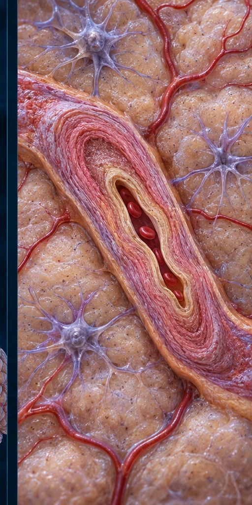
Chronic hypertension can damage vessels. Cerebral amyloid angiopathy and other vascular abnormalities can contribute to hemorrhage. Aneurysmal SAH has a distinct vessel-wall cause. The team investigates the actual event.
Wall-associated plaque, a possible atrial clot source and an intact small artery. Heart chamber geometry is stylized: no precise appendage is identified. The small-artery image cannot establish hypertension, microscopic pathology or future rupture. The text explains the medically supported relationships.
Risk factors that can be addressed
High blood pressure, smoking, diabetes, unhealthy blood lipids, inactivity and some heart conditions increase risk. Medical care and sustainable habits can reduce risk; they cannot guarantee that stroke will never occur.
Risk and personal context
Age, family history and prior stroke/TIA inform risk, and stroke also occurs in younger people. Individual plans account for the event’s cause and other conditions. Symptoms deserve emergency attention even without known risk factors.
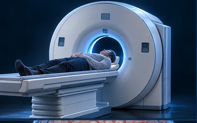
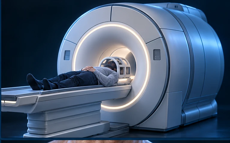
Urgent evaluation · different tests answer different questions
Is there bleeding? Which tissue and vessels are affected?
History, neurological examination and imaging work together. The team asks about symptom onset or last known well, medicines and other conditions; glucose testing also helps identify a stroke mimic. Assessment and treatment planning proceed urgently.
Brain CT
Rapid X-ray cross-sections help identify hemorrhage. Early ischemic changes can be subtle; a normal-looking early CT does not exclude ischemic stroke.
Brain MRI
Magnetic fields and radio waves provide tissue contrasts. Diffusion-weighted MRI can show early ischemic injury; not every patient must wait for MRI before treatment.
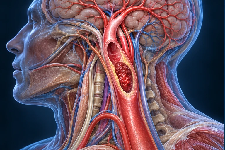
Vessel imaging
CTA, MRA or catheter angiography can locate an occlusion or vascular abnormality. Selected perfusion studies help assess tissue at risk.
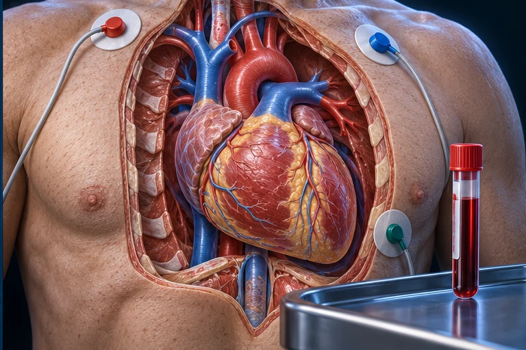
Heart rhythm & blood tests
ECG and monitoring can detect AF. Blood tests assess glucose, clotting and other factors relevant to diagnosis and treatment safety.
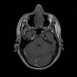
Real MRI stays real
Preserved NLM Visible Human research MRI · public-domain male cadaver specimen, axial sample m_vm1125, source-derived 256 × 256 pixels. This image teaches modality and anatomy; it is not a living patient or a stroke diagnosis example. Enlarging it adds no acquired detail.
The generated equipment and anatomical contexts above explain what each test investigates; they are not scan results. Fine device geometry and electrode positions are illustrative. No generated “clinical imaging” is presented. Courtesy of U.S. National Library of Medicine; no institutional endorsement implied.
Restore flow, or control bleeding. First establish the type.
For eligible ischemic stroke
Intravenous thrombolysis uses alteplase or tenecteplase to dissolve a clot in eligible patients with disabling deficits, usually within 4.5 hours of known symptom onset (or last known well). Selected later or unknown-onset cases may qualify using imaging criteria. Clinicians weigh bleeding risk, imaging, timing and other eligibility factors.
Endovascular thrombectomy retrieves a clot in selected large-vessel occlusions; some eligible patients can benefit up to 24 hours. It is not suitable for every stroke. These are selection windows, not permission to wait or guarantees of recovery. Brain imaging must exclude hemorrhage and assess relevant eligibility; the presence of a clot alone does not make either treatment suitable.
Care may involve close neurological monitoring, controlled blood-pressure management, reversal of anticoagulation where indicated, and assessment of swelling or hydrocephalus. Selected patients need neurosurgical procedures; not every ICH benefits from evacuation.
For aneurysmal SAH, specialists may secure the ruptured aneurysm with an endovascular or surgical approach. This is a subtype-specific pathway, not clot-dissolving treatment. Intensive care and complication surveillance remain important.
One intravenous principle, two views: venous administration and clot breakdown.
1 · Intravenous administration
Alteplase or tenecteplase is given intravenously to eligible patients. After venous administration, medicine circulates through the heart and lungs into the arterial circulation. It can then act on a susceptible clot. The background organs indicate this systemic route; their fine vascular connections are illustrative.
These medicines promote conversion of plasminogen to plasmin, which breaks down the clot’s fibrin scaffold. Restoration of flow is a treatment aim, not a guaranteed result. This image does not represent dosing or a measured before–after sequence.
3 · Mechanical thrombectomy
Specialists reach a selected cerebral large-vessel occlusion through arterial access. This view shows direct contact aspiration only. Stent retrieval is a different technique using an expandable retriever; it is not shown or combined with this catheter.
IV administration and clot dissolution are two views of one treatment principle, distinct from endovascular removal. Both can be considered in an eligible patient; the buttons highlight, rather than select a personal treatment. These are illustrative scales, not exact commercial device designs, operative instructions or a full access route. The magnified catheter is inside an artery; the peripheral IV is inside a vein. All three views concern selected ischemic stroke, not hemorrhage.
Never use this page to choose a medicine or decide you are “too late.” Emergency specialists select treatment. Do not start, stop or combine prescribed medicines without clinical advice.
Assisted task practice with illustrative nervous-system context. The overlay is not a traced motor pathway or proof that a tract has repaired. It is not a home exercise or swallowing-safety instruction.
Restoration and compensation
A coordinated team may work on mobility, daily activities, communication, cognition and swallowing safety. Rehabilitation combines guided task practice with adaptations or assistive strategies. Timing and intensity are tailored to clinical stability and goals, rather than a universal home exercise schedule.
Reassessment can reveal new needs. Mood, fatigue, caregiver training and support affect participation. Recovery varies; improvement may continue over time, but full recovery is not guaranteed.
Prevention follows the cause
Plans address blood pressure, lipids, diabetes, smoking, diet and safe activity. For selected ischemic causes, clinicians choose antiplatelet treatment; AF may require anticoagulation. These are different medicines and are not interchangeable. Prevention after hemorrhage also requires an individualized risk–benefit assessment.
A plan for life after stroke
Follow-up connects the acute team, rehabilitation and primary care. Practical goals can include safer walking, dressing, communication and returning to meaningful activities. Any new sudden warning signs need emergency action, even during recovery.
AF is explained here as a mechanism; no empty AF or future disease page is linked. “Next” above advances to this chapter’s existing recovery section.
MECHANISM QUESTIONS · AFTER THE EMERGENCY MESSAGE
Connect recognition, assessment and recovery.
Why can symptoms prompt action but not choose a medicine?
Sudden neurological change calls for emergency assessment. Similar symptoms can arise from blocked delivery, bleeding or another cause. Brain and vessel assessment help distinguish these possibilities; a recognition picture cannot determine the stroke type.
Does a treatment window mean there is time to wait?
No. Time, imaging, the affected vessels, disabling deficits and bleeding risk influence clinical eligibility. Selected later or unknown-onset cases may still have options; no diagram or clock on this page can decide an individual’s eligibility. Emergency action remains urgent.
Is rehabilitation only about repairing one visible tract?
Recovery can involve relearning skills, adapting tasks and using compensatory strategies with a coordinated team. The assisted-practice illustration supplies a learning context, not evidence that a particular pathway has regrown. Goals and support are reassessed over time.
Original English medical education chapter. Medical evidence check: 10 October 2026; treatment and recognition source recheck: 13 October 2026. Human medical review: Not performed. Physical-phone acceptance: Pending. This is not personal diagnosis or a treatment recommendation.
The existing reference-book study informed topic scope (FB061), not wording or artwork. Official AHA/ASA summaries and available NIH/CDC material support the stated teaching relationships. Direct NINDS pages and the full AIS journal document returned access errors; indexed official text and accessible summaries were used. A full-guideline line-by-line review is not claimed.
Limits: generated vascular branching, exact tissue borders, lesion dimensions, heart chamber geometry, fine microstructure and recovery overlays are illustrative. Recognition scenes are generated, not patient photographs. Equipment illustrations are not scan results. The cellular plate is not microscopy. Precise diagnosis, lesion localization and outcome cannot be inferred from these teaching images. No authentic acute stroke CT, DWI MRI, CTA/MRA case set or validated arterial-territory map has been added. The retained research MRI must not be read as a stroke case. Treatment selection, contraindications, dosing and individual prognosis belong to clinical teams. Video remains future support; no playback or fabricated duration.