A selective tissue environment
Flexible cells can negotiate narrow gaps. Retention and cellular recognition also influence clearance; neither appearance nor one drawing gives a universal age or stiffness threshold.
SPLEEN / THE 30-SECOND PICTURE
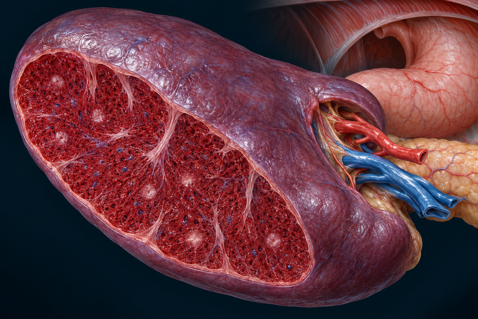
Beside the stomach in the left upper abdomen, a blood-rich organ brings circulation and immune tissue together. Red pulp handles cells; white pulp organises responses to blood-borne material.
The arterial branch approaches the hilum. Smaller routes supply immune regions and the blood-handling pulp. This is not an incoming lymph vessel.
The outline selects a learning region. It does not measure flow, cell activity or immune activation.
Structure and mechanism references: Kenhub · Selected gross splenic anatomy · Histology Guide · MH085 human spleen · Digital Histology · Splenic circulation
01 / CORD TISSUE → SINUS WALL → VASCULAR LUMEN
Locate the open sinus, long lining cells, narrow slit and deforming red cell. Then compare that return route with uptake by a macrophage in the surrounding cord.

In the open route being illustrated, cells enter the cord environment before returning to a sinus. Reticular support and resident cells make this more than an empty space.
The outline selects a learning region. It does not measure flow, cell activity or immune activation.
Flexible cells can negotiate narrow gaps. Retention and cellular recognition also influence clearance; neither appearance nor one drawing gives a universal age or stiffness threshold.
This is not a kidney glomerulus producing a cell-free urinary filtrate. Cells themselves encounter the splenic tissue route.
Compare the renal filtration barrier →Structure and mechanism references: Columbia University · Spleen histology · Digital Histology · Splenic circulation · OpenStax · Erythrocyte structure and recycling · Steiniger and colleagues · 2011 human microcirculation
02 / VESSEL → PERIARTERIOLAR SHEATH → NEIGHBOURING FOLLICLE
Follow the arteriole through its lymphocyte-rich sheath, then look beside it for the follicle. The lighter centre represents one activated context, not every normal nodule.

The periarteriolar lymphoid sheath is associated with an arterial route. It is not the central cavity of a lymph node.
The outline selects a learning region. It does not measure flow, cell activity or immune activation.
Blood-borne material can be captured and presented in a specialised environment. T-cell-rich and B-cell-rich areas support different interactions; the shape or colour of a generated cell does not establish its markers.
Continue with immune recognition →The spleen does not have the lymph-node cortex–medulla plan or its incoming tissue-lymph route. Human boundary compartments also cannot be assumed identical to a mouse’s.
Reconnect with the immune-organ overview →Structure and mechanism references: Histology Guide · MH085 human spleen · Columbia University · Spleen histology · Steiniger · 2015 human and rodent microanatomy · OpenStax · Lymphoid organisation
GO DEEPER / STRUCTURE, CELL HANDLING AND IMMUNE COORDINATION
The spleen occupies the left upper abdomen, close to the stomach and beneath the diaphragm. Its inward-facing visceral surface meets a hilum where vessels enter and leave; its outer surface fits against the diaphragmatic side of the abdominal cavity.
Nearness is not the same as continuity. The pancreatic tail can approach the hilum, and tissue folds connect the spleen with neighbouring structures. The spleen has no digestive duct opening into the stomach or pancreas.
Structure and mechanism references: Kenhub · Selected gross splenic anatomy · Kenhub · Splenic vein course
A connective-tissue capsule encloses the organ. Trabeculae extend inward and support vessels. The broad cutaway makes these supporting strands visible, but they are not complete walls dividing the spleen into identical sealed lobules.
Red and white pulp lie in a connected living tissue environment. This differs from calling one outer shell cortex and an inner core medulla: that plan belongs to other organs, not the splenic pulp.
Structure and mechanism references: Histology Guide · MH085 human spleen · Columbia University · Spleen histology
The splenic artery branches on approaching the hilum. Inside, vessels follow supporting tissue before smaller arterial routes supply the pulp. Some arterial segments have a surrounding lymphocyte-rich sheath; the relationship between a vessel and its tissue is central to understanding white pulp.
After passage through the red-pulp environment, blood is collected in venous sinuses and progressively larger venous channels. The splenic vein leaves at the hilum. A direction cue across the organ is an orientation aid, not a map of every microscopic branch.
Structure and mechanism references: Kenhub · Selected gross splenic anatomy · Digital Histology · Splenic circulation · Histology Guide · MH085 human spleen
Splenic cords contain a supporting reticular network and blood and immune cells. Macrophages occupy this tissue environment. A blood-rich space in a cord is therefore different from a sinus lumen surrounded by endothelial cells.
In an open microcirculatory route, blood enters the cord environment before returning through openings in a sinus wall. Human reconstruction studies address this unusual organisation. The local teaching view follows that route without assigning an exact fraction or tracing every capillary.
Structure and mechanism references: Columbia University · Spleen histology · Digital Histology · Splenic circulation · Steiniger and colleagues · 2011 human microcirculation
A mature red cell is a flexible anucleate cell. Its membrane and supporting cytoskeleton allow deformation. In the red-pulp illustration, the narrowed red cell spans a gap between sinus lining cells; it is not being cut into two pieces.
Mechanical passage is part of selection, not the whole explanation. Reduced deformability and surface changes can contribute to retention and clearance. Colour in an illustration does not by itself establish a cell’s age, measured stiffness or disease.
Structure and mechanism references: OpenStax · Erythrocyte structure and recycling · Columbia University · Spleen histology · Merck Manual Professional · Normal splenic roles
Some cells return to the sinus lumen and circulation. Macrophages can take up retained or altered cells and cellular material. These are distinct processes, shown at separate sites rather than as one compulsory fate for every passing red cell.
The spleen is not the only place where red cells are cleared. Macrophages in other organs also participate. A local image explains one important environment; it does not measure the proportion of all clearance performed there.
Structure and mechanism references: OpenStax · Erythrocyte structure and recycling · Merck Manual Professional · Normal splenic roles · Histology Guide · MH085 human spleen
When a macrophage breaks down a red cell, haemoglobin does not simply become a replacement red cell. Globin can supply amino acids. Iron can enter storage or return to the circulating iron pool, including transport associated with transferrin toward marrow.
The remaining haem portion contributes to pigment processing, with bilirubin carried toward the liver. This connects splenic clearance with hepatic handling and biliary elimination. The spleen is not a bile-making organ and has no bile duct to the gallbladder.
Structure and mechanism references: OpenStax · Erythrocyte structure and recycling · Kenhub · Selected gross splenic anatomy
The periarteriolar lymphoid sheath, or PALS, is a T-cell-rich region around an arteriole. A B-cell follicle lies alongside this route. An active follicle may develop a germinal centre, so the named central arteriole can appear off to one side of the follicle.
These are functional tissue regions, not cell identities diagnosed from purple colour alone. Antigen presentation, cell cooperation and regulated activation connect blood-borne material with an adaptive response. A photograph or generated cell profile cannot establish a receptor’s specificity.
Structure and mechanism references: Histology Guide · MH085 human spleen · Columbia University · Spleen histology · OpenStax · Lymphoid organisation
The spleen provides a setting in which mature lymphocytes meet relevant signals, become activated and expand. Antibody-secreting plasma cells arise from activated B-cell responses. T-cell and B-cell contributions are complementary, rather than interchangeable versions of one cell.
Initial lymphocyte development has other organ contexts: bone marrow and thymus are primary lymphoid organs. Calling the spleen secondary does not make it unimportant; it distinguishes a different stage of immune organisation.
Structure and mechanism references: OpenStax · Lymphoid organisation · Merck Manual Professional · Normal splenic roles · Columbia University · Spleen histology
The boundary between white and red pulp contains specialised cell and vascular relationships. Human and rodent compartments differ. A textbook-style ring labelled as a universal marginal sinus would hide those differences.
The white-pulp image therefore identifies the arteriole, surrounding sheath, adjacent follicular region and neighbouring red pulp. It does not claim to resolve every human marginal or perifollicular subdivision. Compare the linked human specimen before drawing conclusions about exact boundaries.
Structure and mechanism references: Steiniger · 2015 human and rodent microanatomy · Histology Guide · MH085 human spleen · Digital Histology · Red and white pulp
The splenic vein courses toward the portal system behind the pancreatic body and tail. That venous pathway is different from bile travelling out of the liver. Existing portal-route lessons show the wider abdominal setting.
Regional lymphatic drainage travels toward hilar and pancreaticosplenic nodes and onward toward coeliac nodes. This does not make the spleen a node receiving the body’s incoming lymph. Arterial-associated autonomic fibres also reach the organ; these illustrations do not certify fine neural branching or a direct human parasympathetic route.
Structure and mechanism references: Kenhub · Selected gross splenic anatomy · Kenhub · Splenic vein course · Histology Guide · MH085 human spleen
The spleen also participates in platelet distribution and hosts changing immune-cell populations. Storage and cellular traffic are normal roles, but their size and dynamics depend on the biological setting; a fixed reservoir percentage is not necessary to understand this chapter.
During development, blood-cell production has organ roles that differ from the usual adult marrow-centred pattern. This normal chapter does not use fetal physiology or a large animal spleen’s contraction as evidence that a human spleen supplies a guaranteed rescue volume.
Structure and mechanism references: OpenStax · Lymphoid organisation · Kenhub · Selected gross splenic anatomy · Merck Manual Professional · Normal splenic roles
The external MH085 reference is a human H&E section. It can show capsule, tissue regions and vessel profiles. Section direction, stain and collapse influence what is visible; a pale circle is not automatically a complete three-dimensional follicle.
Our illustrations expose selected anatomy at several scales. They are original generated teaching artwork, not patient scans, microscopy or a reconstruction from that specimen. Their labels name broad verified relationships without certifying every fine generated cell, strand or opening.
Structure and mechanism references: Histology Guide · MH085 human spleen · Columbia University · Spleen histology · Digital Histology · Red and white pulp
REAL HUMAN TISSUE / A DIFFERENT KIND OF EVIDENCE
MH085 is identified by Histology Guide as human spleen stained with H&E. Begin at the capsule, find pale and dense lymphoid regions, and compare them with the blood-rich surrounding pulp. A section cuts across structures at different angles.
These are external specimen references. Their slide pixels are not redistributed here. Our generated illustrations are not microscopy, immune-marker stains or reconstructions derived from those patients.
Structure and mechanism references: Histology Guide · MH085 human spleen · Columbia University · Spleen histology
No. The main incoming route is arterial blood. This differs from a lymph node receiving lymph from a tissue drainage area.
No. Return to a sinus and macrophage uptake are distinct outcomes at different sites.
No. The arteriole is associated with PALS, beside a B-cell follicle. The follicle can make the arteriole appear eccentric.
No. Iron and globin recovery and pigment processing have different onward routes. Hepatic processing belongs to the liver and biliary lessons.
Targeted normal structure and function checks: 20 October 2026. Existing FB102 book research guided the organ’s place in the body-wide curriculum; independent sources support the tissue and mechanism explanations. No source text, textbook composition, institutional slide or atlas figure was copied.
Original text-free generated teaching bases, editable semantic names and stable anatomical targets are separate layers. The normal anatomy does not select treatment or diagnose symptoms. Fine generated anatomy, physician review, actual physical-phone use and independent post-publication review are not certified by these checks.