An internal space can still face an epithelial boundary
An airway, gut lumen or bladder cavity is separated from supporting tissue by a living lining. An internal-looking space is not automatically part of the connective-tissue compartment. Locate the surface first, then ask what can cross it.
Skin faces the exterior, respiratory lining faces an airway, and urothelium faces urine. These pictures share an orientation convention, not one anatomical location. Glandular cells can arrange around secretory spaces; endocrine organisation needs its own context.
Structure and function: Histology Guide · Epithelial organisation · OpenStax · Epithelial tissue, critically compared
Apical and basal describe relationships, not gravity
In these covering and lining examples, the apical domain faces the free space. Lateral membranes face neighbours. The basal domain faces the supporting extracellular interface. Turning a picture upside down does not exchange these biological identities.
Different membrane domains can carry different transport, adhesion and secretion machinery. Maintaining a sheet therefore means preserving the right activities on the right faces, as well as keeping cells together. Rendered texture does not identify those molecules.
Structure and function: Apical–basal polarity and epithelial function · Pickett, Naturale & Feldman · Tissue context and polarity
Count layers by cells and their attachment
A simple epithelium has one layer whose cells contact the basal supporting boundary. In a stratified sheet, only the basal population reaches that boundary. Nuclear rows alone are unreliable because a section crosses different parts of different cells.
Pseudostratified epithelium looks layered because nuclei occupy different heights, although cells share one basal attachment level. Some basal cells do not reach the free surface. This must not be drawn as several complete stacked cell tiers.
Structure and function: Histology Guide · Epithelial organisation · OpenStax · Epithelial tissue, critically compared
The exposed population helps name a stratified sheet
Squamous, cuboidal and columnar describe flattened, roughly equally tall-and-wide, and taller-than-wide profiles. In a stratified tissue, the surface population supplies this part of the name. Cuboidal basal cells do not make a squamous surface stratified cuboidal.
Cells are not perfect solids. Their outlines vary with tissue state and section direction. Natural irregularities in an atlas are not measurements of cell geometry.
Structure and function: Histology Guide · Epithelial organisation
Thinness serves different organs in different ways
The new squamous preparation depicts mesothelium on connective support: a thin living surface contributing to a smooth serosal interface. Endothelium belongs to vessels. Similar simple squamous profiles do not make these locations interchangeable.
Alveolar exchange, a vascular interface and a moving serosal surface need different supporting structures. The linked human kidney field shows the parietal lining of Bowman’s capsule; that lining is not the whole glomerular filtration barrier.
Structure and function: Histology Guide · Epithelial organisation · Histology Guide · MH016 human kidney field · OpenStax · Epithelial tissue, critically compared
A short epithelial profile can support substantial transport
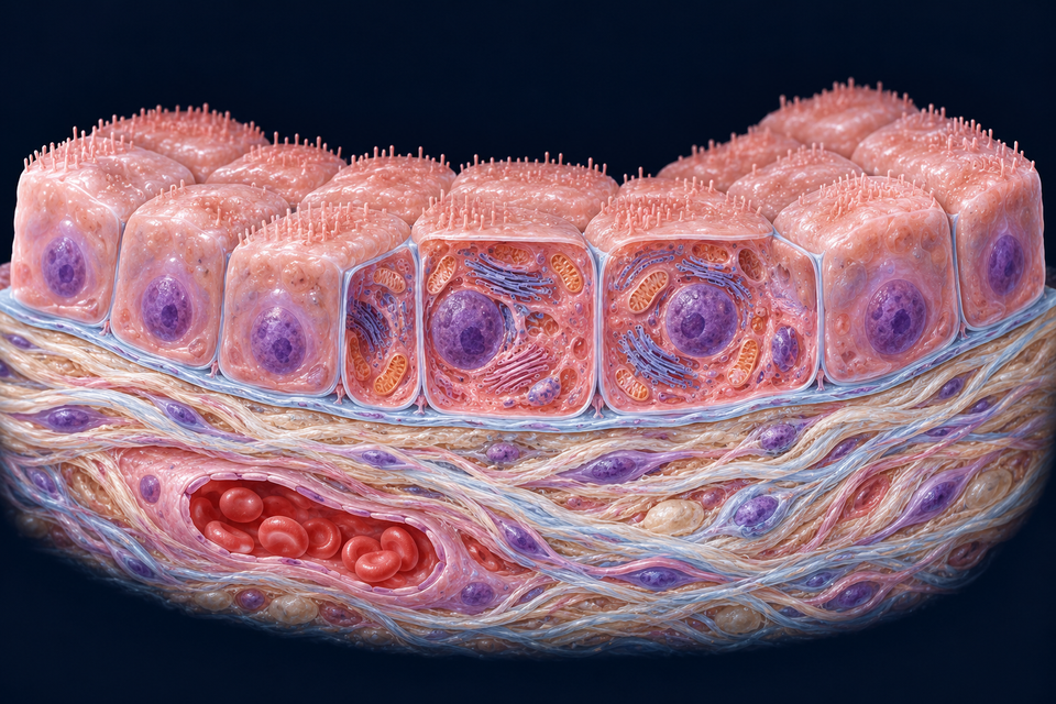
Many small glandular ducts and renal tubular regions contain cuboidal cells. A short profile does not imply an inactive surface. Membrane organisation and cytoplasm can support ion movement, recovery or secretion.
The opening view is generic. Its organelles and short surface projections do not establish a particular kidney segment or gland. Different ducts modify different secretions; cell shape alone does not predict the fluid leaving them.
Structure and function: Histology Guide · Epithelial organisation · Apical–basal polarity and epithelial function
A tall absorptive cell has more than one working surface
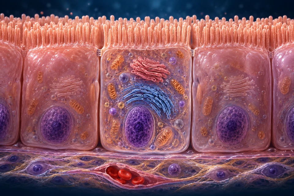
The retained intestinal view places microvilli at the lumen-facing edge and a tissue-facing domain below. The two faces perform different parts of coordinated transfer. Apical uptake alone does not prove that material has reached blood or lymph.
A villus is a multicellular mucosal projection containing supporting tissue. Microvilli are much smaller projections of individual cells. This opened enterocyte complements the intestinal lesson rather than mapping every transporter or secretory cell.
Structure and function: Histology Guide · Epithelial organisation · NCBI Bookshelf · Cell–cell interactions
Staggered cells and motile cilia cooperate at a wet surface
The respiratory example retains the qualified nasal mucosa. Tall ciliated profiles, secretory examples and a small basal region contribute to pseudostratified organisation. Mucus and surface fluid lie above the cells; vessels remain in separate support below.
Cilia help carry surface material toward clearance. That movement differs from air reversing during breathing and from olfactory sensory detection. The latter uses another specialised tissue organisation and neural pathway.
Structure and function: Histology Guide · MH134 human respiratory lining · Histology Guide · Epithelial organisation
Several layers preserve a surface while cells change
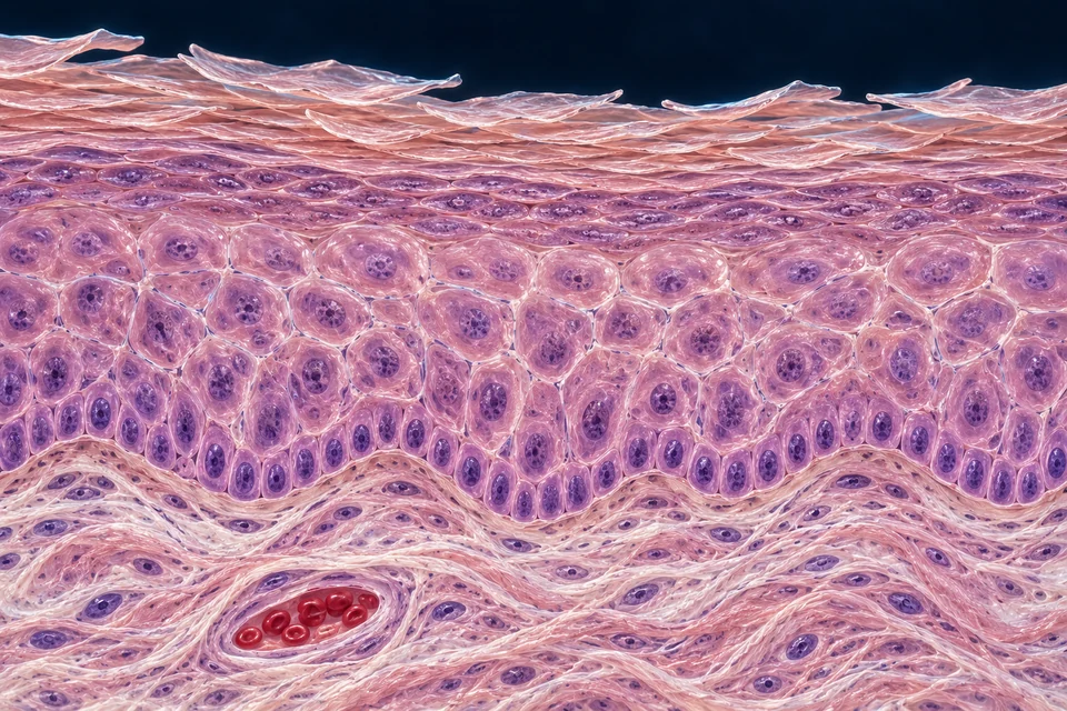
The retained skin view places basal and viable layers beneath a cornified surface. Newer basal cells and the outer flattened population have different roles. A capillary belongs below the epithelial–dermal interface, not inside the epidermis.
Not every stratified squamous surface is keratinized like skin. Oral, oesophageal and corneal epithelia have region-specific non-keratinized arrangements. Corneal transparency and renewal need their own organ context; the cornea is not a sheet of skin covering an eye.
Structure and function: Histology Guide · Epithelial organisation · OpenStax · Epithelial tissue, critically compared
A distensible surface remains a specialised barrier
Urothelium combines basal, intermediate and surface umbrella populations. Its surface cells and membrane organisation adapt to changing wall shape while maintaining a restrictive interface. Flattening during filling does not turn it into ordinary stratified squamous epithelium.
The picture is a structural snapshot, not a recording of filling or molecular plaques. Underlying muscle produces organ force; it is not the urine-contacting barrier. Resistance, sensation and movement are related but distinct functions.
Structure and function: The urothelium: life in a liquid environment · Histology Guide · Epithelial organisation
The supporting boundary is extracellular, not another cell tier
The basement-membrane region is specialised extracellular matrix associated with the epithelial base. It supports attachment and spatial organisation. Its thin pictured interface does not independently resolve the molecular layers.
Basal support and lateral cell adhesion are different relationships. A matrix attachment does not join two neighbours. Composition and appearance vary among tissues and preparations, so a coloured rim cannot certify an identical molecular structure everywhere.
Structure and function: Histology Guide · Epithelial organisation · Pickett, Naturale & Feldman · Tissue context and polarity · OpenStax · Epithelial tissue, critically compared
Avascular does not mean unsupported
The ordinary surface sheets illustrated here have no vessels within their epithelial layers. Circulation in connective support supplies exchange across intervening tissue spaces. In a thick stratified sheet, the surface is farther from that supporting circulation.
An absorptive lumen is still not a blood vessel. Cell nutrition does not follow one identical route in every epithelium. Epithelium, matrix, fluid and circulation work together; the cues here do not measure diffusion distance or flow.
Structure and function: Histology Guide · Epithelial organisation · OpenStax · Epithelial tissue, critically compared
Seal, hold and communicate are different connection jobs
Tight junctions regulate the between-cell route and help separate membrane domains. Adherens junctions and desmosomes supply mechanical links between neighbours. Gap junctions couple adjacent cytoplasms for selected small signals and ions; they are not lumen-to-tissue holes.
Appropriate basal attachments, including hemidesmosomal anchorage, connect a cell to matrix. The primary picture locates a contact region without assigning a named protein complex to unverified texture. Different tissues do not use one universal junction distribution.
Structure and function: NCBI Bookshelf · Cell–cell interactions · Apical–basal polarity and epithelial function · OpenStax · Epithelial tissue, critically compared
Through a cell and between cells are different pathways
A transcellular route crosses membrane domains and the cell interior. A paracellular route follows the extracellular path between neighbours under junctional control. Their contributions depend on the tissue and substance.
A tight junction is not a universal zero-permeability weld. Selectivity and regulation matter. The route cues distinguish locations; they do not show measured rates, open pores or a recommendation to increase permeability.
Structure and function: NCBI Bookshelf · Cell–cell interactions · Apical–basal polarity and epithelial function
Microvilli and motile cilia solve different problems
Microvilli enlarge an apical membrane surface for absorption or other local work. Motile cilia organise movement over a surface. Compare the intestinal brush border and respiratory ciliary field by function as well as appearance.
Not every visible projection is one of these structures, and not every cilium is motile. The generic cuboidal view’s short projections are not assigned to a renal segment. Molecular supports and beat mechanics require evidence beyond rendered outlines.
Structure and function: Histology Guide · Epithelial organisation · Histology Guide · MH134 human respiratory lining · OpenStax · Epithelial tissue, critically compared
A gland organises an epithelial output
An exocrine output reaches a surface or lumen, often through a duct. Secretory end pieces and downstream duct cells can do different jobs. A goblet cell is a small secretory example within a lining; a salivary gland arranges many epithelial units with support.
Endocrine output reaches the internal environment through neighbouring tissue and circulation, rather than an excretory duct to a surface. This links to the existing Mouth and Endocrine lessons. Blood does not normally empty into an exocrine duct.
Structure and function: OpenStax · Epithelial tissue, critically compared · Apical–basal polarity and epithelial function
A boundary renews without one universal timetable
Regional progenitors contribute to maintenance in different ways in skin, intestine, airway and other tissues. Skin maturation changes cells approaching the outer surface. A quiet urothelial snapshot does not depict the same rapid renewal pattern as an intestinal lining.
No single period, cell-travel direction or layer count applies to all epithelia. This normal lesson does not assess a wound, grade a biopsy or recommend treatment.
Structure and function: Histology Guide · Epithelial organisation · Pickett, Naturale & Feldman · Tissue context and polarity · OpenStax · Epithelial tissue, critically compared
A section is a view through tissue
A section can cross a nucleus, miss it or meet a sheet obliquely. Cell proportions and visible nuclear levels need anatomical context. Fixed H&E cannot demonstrate live ciliary motion or directly measure selective transport.
MH016 is mixed-species: kidney and ovary are identified as human, trachea and small intestine as monkey. The link is specifically to the human kidney discussion. The generated preparations here remain illustrations, not additional specimen evidence.
Structure and function: Histology Guide · MH016 human kidney field · Histology Guide · Epithelial organisation