Relay is active processing
Thalamic nuclei connect with particular cortical regions. They participate in sensory, motor and state-related circuits; they are more than a passive junction.
Evidence: Central processing · OpenStax
Nervous system · chapter
The brain brings sensation, movement, memory and the body’s internal needs into a changing pattern of activity.
Start with its folded surface. Then look inward to the tissue, fluid spaces and connected circuits that make coordinated behaviour possible.
See the connected brain →Left lateral view
Anterior ← → Posterior
Colours distinguish regions; they are not natural tissue colours.
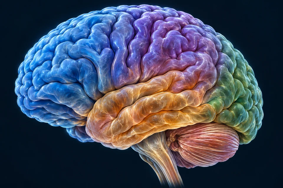
30-second orientation · visible regions, connected jobs
A medial section reveals relationships hidden behind the outer folds. The frontal end is left; the cerebellum and back of the brain are right.
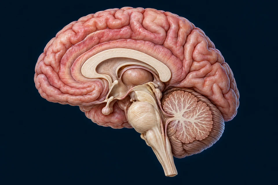
Different regions cooperateInformation reaches the brain, is processed across connected networks, and influences bodily responses. Feedback keeps changing the activity; a lobe or nucleus is not an isolated box with one job.
Cyan rings locate contexts only. The three phrases summarise relationships, not exact axon trajectories or a universal serial processing chain. Follow sensation and action →
Regional and functional sources · OpenStax · checked 17 October 2026.
Thalamic nuclei connect with particular cortical regions. They participate in sensory, motor and state-related circuits; they are more than a passive junction.
Evidence: Central processing · OpenStax
The caudate, putamen and globus pallidus belong to the basal nuclei. Together with wider circuits, they help regulate movement. They sit away from the midline and are not individually traced in this plate.
Evidence: Anatomy & Physiology · OpenStax
The internal capsule is a concentrated white-matter route between deep gray regions. Descending motor fibres continue through the brainstem; many cross in the medulla before reaching spinal circuits.
Evidence: Motor routes · OpenStax
Generated gross anatomical context. Fine nuclei, fibre trajectories, exact cut plane and individual boundaries remain illustrative. The hippocampus is not inserted into this midline view.
FROM THE PREPARATION TO THE SOURCE MODEL
Keep the chapter preparations alongside the existing adult male reference. Select a named source structure, inspect its location or use text view, then return to the corresponding lesson. These are related teaching links, not newly approved anatomical representations.
Locate the lobes, then compare their connected functions.
Gross regions do not identify exact functional borders or cortical cell layers.
Connect the deep anatomy with processing and internal regulation.
These surfaces do not resolve individual nuclei, SCN cells or endocrine pathways.
Use the chapter to understand loops that influence action.
No direct or indirect circuit, neurotransmitter release or fine nucleus subdivision is traced.
Compare a named commissure with the three connection families.
The source shape is not a measured fibre bundle or a tractography image.
Connect the source location with memory and learning mechanisms.
This is not a memory store diagram, cellular layer preparation or synaptic recording.
Distinguish midbrain, pons and medulla in their wider context.
Cranial nerve nuclei and exact ascending or descending pathways are not certified.
Study coordination alongside the chapter tissue and feedback explanations.
The model does not show Purkinje cells, cortical layers or circuit activity.
Compare fluid spaces with the surrounding solid tissues.
Cavity surfaces represent spaces, not solid brain tissue, fluid flow or a patient scan.
Native shapes and coordinates are retained. Ventricular surfaces locate fluid spaces, not solid nervous tissue. The chapter preparations explain cellular organisation and mechanisms beyond these gross surfaces. Source credits remain available in the viewer.
From surface to depth
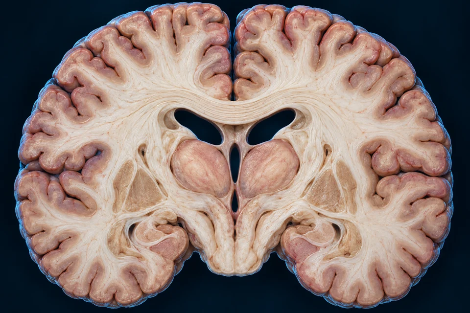
A coronal section cuts across both hemispheres. Gray matter contains many cell bodies, dendrites and synapses; white matter contains many axons, including myelinated fibres. Deep nuclei are gray matter within this interior. The lateral ventricles are CSF-filled cavities, not solid tissue; their full arrangement is shown in the next illustrated topic.
Association fibres link regions in one hemisphere. Commissural fibres cross between hemispheres. Projection fibres connect the cortex with deeper centres and spinal routes.
Gyri are raised folds; sulci are grooves. The central sulcus separates frontal from parietal cortex. The lateral sulcus lies above the temporal lobe. Boundaries help orientation; functions extend across them.
Frontal coronal view; a schematic anatomical illustration, not a measured specimen. Fine fibre paths and individual deep nuclei are not traced.
Evidence: Regional anatomy
Regions work together
Reaching for a cup combines visual location, a movement plan, descending commands and continuous feedback. The lobes are landmarks, not isolated boxes with one job each.
Loops with cortex and thalamus help select and regulate actions.
Compares movement-related information and feedback, helping adjust timing and accuracy.
Midbrain, pons and medulla carry pathways and house centres involved in breathing, circulation and arousal.
Ascending pathways convey sensory information; descending pathways influence movement. Some reflex processing occurs in spinal circuits.
Connection map supports the anatomy; arrows summarize relationships rather than exact axon trajectories.
Evidence: Regional anatomy
A second surface view · not another lateral picture
Looking from above makes the midline visible. The connecting corpus callosum is below this surface, so it belongs in the section view rather than on top of the brain.

A gyrus is a raised fold; a sulcus is a groove. The central sulcus separates frontal from parietal cortex, while the longitudinal fissure separates hemispheres. The generated folds here are not an exact individual sulcal atlas, so no precise functional border is traced on them.
Lower-limb motor and sensory representations extend onto cortex facing the midline. Much of primary visual cortex also lies medially, farther back. A lateral image cannot reveal every area; combining top, side and section views is more informative than repeating one angle.
Folding accommodates a large cortical sheet inside the skull. Gray matter contains local cellular circuits; underlying white matter connects regions. A lobe name describes a broad anatomical territory, while a task such as reading depends on communication across several territories.
Evidence: OpenStax
Generated gross anatomy, not a scan. Hemisphere separation and orientation are the teaching scope. Individual gyri, sulci, surface vessels and functional boundaries remain illustrative; anatomical left/right is not inferred from an unmarked scan convention.
Regional anatomy · gray nuclei and white connections
Compare cortex, white matter and deep nuclei in two complementary planes. Each selected view has its own orientation key.
Axial view · anterior at the top

Projection · between levelsAscending and descending connections link cortex with deeper centres, brainstem and spinal routes. Here the internal capsule is the visible white corridor between deep gray regions; it is not another nucleus.
This plane adds information the midline cannot show. Anterior is at the top. The labels identify broad regions on one side; paired structures are also present on the other.
Use the same anatomical views to distinguish a white-matter bridge, a deep corridor and connections within one hemisphere. The buttons change the view and explanation; they do not identify individual axons.
The caudate and putamen form the dorsal striatum. The putamen and globus pallidus form the lentiform region. These names group structures in different ways; the white internal capsule is not another nucleus.
The capsule’s anterior limb lies between caudate and lentiform tissue; its posterior limb lies between thalamus and lentiform tissue. Fibres spread above it as the corona radiata. Ascending sensory and descending motor connections share this crowded corridor.
A small injury in this concentrated white matter can affect several connections. Location matters as well as size. This teaching section cannot identify an individual lesion or predict its effects.
Evidence: NCBI / NLM · OpenStax
Basal-nuclear output normally restrains parts of the thalamic motor circuit. Changing that inhibition helps permit selected actions and suppress competing activity. Dopamine modifies the circuit; it is not a simple instruction to contract a muscle.
Evidence: Published anatomy review
Connect neighbouring or distant cortical regions, allowing information from one area to inform another.
The corpus callosum links the hemispheres. Communication between sides helps combine information and coordinate responses.
Connect cortex with deeper centres, brainstem and spinal routes. The internal capsule is a compact part of these connections.
Evidence: OpenStax · NCBI / NLM
Generated regional anatomy, checked for the stated neighbouring relationships. Individual nuclear boundaries, capsule subdivisions and fine fibres remain illustrative; this is neither MRI nor diffusion tractography.
Regional magnification · tissue to feedback

The brainstem contains pathways and local nuclei. Midbrain circuits contribute to orienting and eye movements; pontine connections link cerebral and cerebellar activity. Medullary networks help regulate breathing and circulation. Many cranial nerves connect here, but the olfactory and optic pathways are not brainstem nerves.
Many corticospinal axons pass through the internal capsule, midbrain and pons, then cross near the lower medulla. They influence opposite-side spinal motor circuits. This describes a major route, not a rule that every sensory or motor connection crosses in the same place.
Evidence: OpenStax
Information about an intended action and feedback from the body arrive through several routes. Cerebellar processing helps correct timing and accuracy. Its output influences other motor centres rather than directly commanding a skeletal muscle.
Evidence: NCBI / NLM
Evidence: OpenStax

Purkinje cells provide inhibitory output from the cerebellar cortex, usually to deep cerebellar nuclei. Those nuclei also receive incoming signals and send outputs onward. A branch in the arbor vitae is white matter, not itself a Purkinje-cell layer.
Evidence: NCBI / NLM · NCBI / NLM
Supporting feedback model, not a computational simulation. Individual deep nuclei and exact peduncular pathways are not traced in the generated plate.
Fluid cavities in their anatomical setting
Cyan represents fluid-filled spaces, not blue brain tissue. Translucent surrounding anatomy lets the main cavities remain visible.
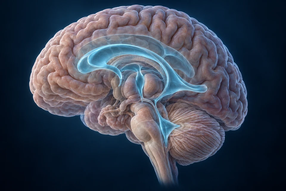
Lateral cavities lie within the hemispheres; the third is central. The aqueduct passes through the midbrain to the fourth, whose floor is the back of the pons and upper medulla.
Small openings are not reliably resolved in the generated plate. The separate route below names the interventricular foramina and fourth-ventricle apertures without pretending to show their exact shapes.
Gross cavity orientation is illustrative; tiny openings, choroid plexus and exact ventricular horns are not individually verified in this rendering. The source-based flow sequence below supplies the teaching connections.
Evidence: Anatomy & Physiology · OpenStax
Protection & a controlled environment
Skull, membranes and cerebrospinal fluid support the brain in different ways. The fluid spaces connect; the protective layers are not interchangeable.

Choroid plexus epithelium secretes most CSF into the ventricles. This is regulated secretion across a blood–CSF barrier, not simply blood leaking into a cavity.
Return has several routes. Arachnoid villi and granulations interface with dural venous sinuses, providing a route toward venous blood. Lymphatic-associated pathways also contribute to outflow; the relative contributions are an active research question.
CSF provides buoyancy and a fluid environment. Motion is pulsatile, and this route diagram is not a steady-flow simulation. Narrowing an outlet can disturb fluid balance; enlarged spaces alone do not establish a diagnosis.
Evidence: Protection and circulation · CSF system review · Figure 1
Cavity → secreting tissue → fluid balance
The ventricular plate shows where CSF travels. This magnification explains the tissue interface that controls much of its production.

Choroid plexuses project into the lateral, third and fourth ventricles. Each vascular projection has a connective-tissue core covered by specialized epithelial cells. This enlarged example shows one projection, not the shape of the entire plexus.
Evidence: Published neuroscience review
Transporters and pumps move ions across the epithelium; associated water movement contributes to CSF secretion. Fenestrated plexus capillaries allow exchange with the core, but epithelial tight junctions restrict passage between covering cells. The barrier is at that epithelium, unlike the endothelial barrier in ordinary brain capillaries.
Evidence: NCBI / NLM · Published neuroscience review
CSF provides buoyancy and helps maintain the chemical environment around the CNS. Production, movement and outflow interact. The route is pulsatile rather than a conveyor belt, and neither this image nor the route diagram measures a production rate or a pressure.
Evidence: Published neuroscience review
Superficial and deep venous systems drain the brain toward dural venous sinuses and ultimately the internal jugular veins. A dural sinus is a blood channel within the dura; it is not a ventricle or the fluid-filled subarachnoid space.
Evidence: NCBI / NLM
Arachnoid projections provide a classical venous interface. Evidence also supports outflow associated with cranial and spinal nerves and meningeal lymphatic vessels. Exact connections and the relative contribution of these routes in humans remain under study; a single arrow to a sinus is an incomplete model.
Evidence: Published neuroscience review · Published neuroscience review
Ordinary ventricular ependyma, specialized plexus epithelium and meningeal barriers are not interchangeable. The capillary seen here supplies the plexus core; it does not discharge red blood cells into CSF.
Evidence: NCBI / NLM · Published neuroscience review
Generated tissue illustration, not histology. The covering/core/vessel/fluid relationship is checked at educational scope. Fine junctions, fenestrations, cell shapes and counts, transport proteins and physical scale are not independently verified in the raster. The controlled diagram and explanations carry those bounded physiological relationships.
Metabolic support
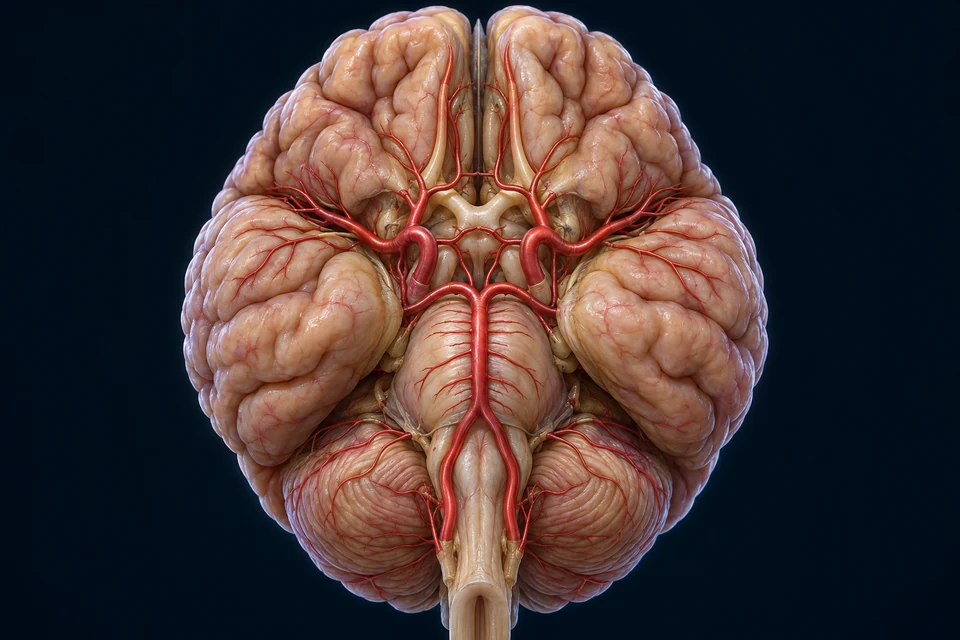
Inferior view; anterior at the top. Major approaches are illustrative anatomical context. Tiny arterial branches, exact ring anatomy and individual vascular territories are not independently verified in the generated image.

The anterior communicating artery (ACom) joins the anterior cerebral arteries (ACA). Each posterior communicating artery (PCom) links a carotid junction with a posterior cerebral artery (PCA). Middle cerebral arteries (MCA) lead outward from the carotid approaches.
The arterial pattern varies between people. A drawn ring is not proof of a complete or effective collateral pathway in an individual.
Evidence: Circle of Willis · NCBI Bookshelf
Anterior cerebral supply includes much of the medial frontal and parietal cortex, where lower-limb representation extends.
Middle cerebral branches supply extensive lateral cortex and contribute deep branches. A single surface label does not show the full territory.
Posterior cerebral supply includes occipital and inferior temporal regions. Territories overlap and vary; this is broad orientation, not an individual vascular map.
Evidence: Protection and circulation
Connecting vessels at the brain’s base form the arterial circle. Its anatomy varies, so it does not guarantee adequate backup supply.
The endothelial barrier regulates exchange. Transport systems supply needed molecules; astrocytes and other cells help maintain the vascular environment. The barrier is selective, not an absolute seal.
This barrier is located at the plexus epithelium. It differs from the endothelial blood–brain barrier and helps control CSF composition.
Original route and interface maps; no exact vessel course or measured transport rate is represented.
Evidence: Protection and circulation · CSF system review · Figure 1
An organ needs a controlled cellular environment

In the capillary above, blood brings oxygen and metabolic substrates to nearby living tissue. Glucose is a major fuel for the adult brain under usual conditions; it is not the only possible fuel. Its metabolism, including oxygen-dependent mitochondrial processes, supports ATP production.
ATP powers work such as the sodium–potassium pump, helping maintain the ion gradients needed for repeated electrical activity. The pump is different from the fast voltage-gated channels that produce an action potential. Interruption of supply can compromise energy availability and ion balance; the effects depend on the circumstances, rather than following one fixed timetable in every cell.
Evidence: Brain energy-metabolism review · OpenStax
Follow the connection: gradients and electrical signals · how cells make ATP.
Endothelial tight junctions restrict passage between cells. Selected transport routes move substances through cells. Pericytes, basement membrane and astrocytes help maintain this interface; astrocyte endfeet do not replace the endothelial seal.
Evidence: Published anatomy review
Oligodendrocytes form sheaths around CNS axons; one cell can support several axon segments. In peripheral nerves, a myelinating Schwann cell supplies one segment. Nodes remain exposed, allowing the electrical signal to be regenerated along the axon.
Astrocytes participate in ion and transmitter balance around neurons. Microglia are resident immune cells. Ependymal cells line ventricles; the specialized choroid-plexus epithelium forms a distinct secreting interface.
Arteries deliver blood to tissue capillaries; veins return it toward dural venous sinuses. CSF outflow interacts with venous and lymphatic routes. A venous sinus, a cerebral vein and a CSF cavity are different spaces.
Evidence: OpenStax · Published anatomy review
Generated explanatory magnification, not histology or electron microscopy. Colours, relative sizes and membrane fine detail are illustrative. The controlled comparison carries the verified barrier distinction.
Cellular scale
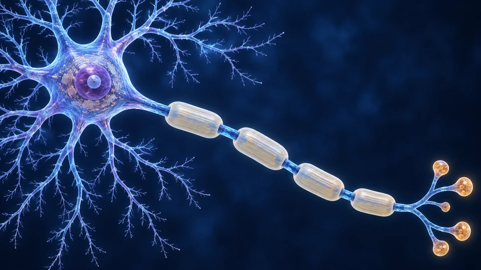
Form myelin around CNS axons. One cell can contribute sheaths to several axons.
Help regulate the extracellular chemical environment and support synaptic and vascular function.
Provide immune surveillance and participate in tissue responses and debris removal.
Line ventricular surfaces. Specialized plexus epithelial cells participate in CSF secretion.
Generated explanatory cell, not microscopy. Organelles, proportions and membrane textures are illustrative; colours do not establish cell identity.
Evidence: Nervous tissue
Electrical along an axon · chemical across a gap
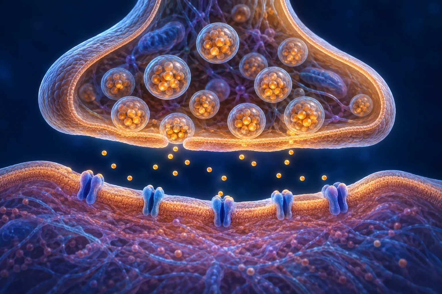
An arriving action potential opens calcium channels. Calcium entry promotes vesicle fusion and transmitter release.
Transmitter crosses the extracellular gap and binds receptors. Receptors change the receiving cell’s activity; effects can be excitatory or inhibitory.
Many small inputs combine over time and across a cell. Their balance influences whether the axon reaches threshold. Transmitter is removed by mechanisms such as uptake or enzymatic breakdown; it does not travel the full axon as the electrical signal.
Resting membrane: Ion gradients and selective membrane permeability establish a voltage across the membrane.
Resting membrane: Ion gradients and selective membrane permeability establish a voltage across the membrane.
Threshold & rise: Sufficient depolarization opens voltage-gated sodium channels. Sodium entry drives the rising phase.
Recovery: Sodium-channel inactivation and potassium movement repolarize the membrane; a refractory period limits immediate refiring.
Propagation: Local currents bring the next axon region to threshold. With myelin, current spreads between nodes and action potentials regenerate at the gaps.
Enlarged generated illustration, not a micrograph. Vesicle counts, molecular shapes and gap dimensions are illustrative; the two cell membranes remain separate.
Evidence: Electrical activity · Synaptic communication
The visible mechanism behind the cell illustration
Excitatory inputs often move membrane potential toward threshold; inhibitory inputs can oppose firing or shunt incoming current. Inputs combine across sites and time. A larger input can change firing frequency, but does not create a proportionally larger action potential.
Evidence: OpenStax
Voltage-gated sodium-channel activation drives the rising phase; channel inactivation and potassium currents help recovery. Myelin reduces current loss between nodes. Current spreads to the next exposed node, where another action potential is generated; the event does not physically leap through empty space.
Evidence: OpenStax
A different region · a different cut
Opening the medial temporal region reveals structures hidden inside the lobe. This oblique view is not the midline section shown earlier.
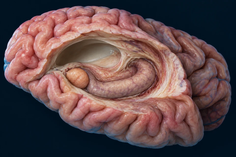
Hippocampal circuits help organize new experiences into memories. Learning and recall also depend on distributed cortical connections. Emotional significance can influence what is learned and remembered.
“Limbic” describes associated structures and connections, not a separate brain acting alone. Memory, motivation and body regulation interact with perception, attention and cortical activity.
Holding a room number while finding a door involves temporary maintenance and active use of information. Attention and frontoparietal networks matter; this is not just a smaller long-term storage bin.
Episodic memory concerns an experience in its setting; semantic memory concerns facts and meanings. Medial temporal structures are important for establishing many new declarative memories, with wider cortical networks contributing to later use.
Improving a practiced movement can involve basal-nuclear and cerebellar circuits without consciously recalling each practice session. Emotional conditioning and familiarity also differ from deliberately recounting an event.
Evidence: OpenStax · Oxford cognitive neuroscience
Plasticity includes changes in how synapses respond and how circuits are recruited. The hippocampus helps relate elements of a new experience; it is not the sole location of every established memory. Consolidation has cellular and wider-network timescales, and retrieval is reconstructive rather than a perfect replay.
Evidence: OpenStax · Oxford cognitive neuroscience
Amygdala-associated circuits help attach significance to events and influence learning. An intense memory can feel vivid without every detail being accurate. Attention, context and later information all affect recall; the amygdala is not a universal emotion switch.
Evidence: OpenStax
This names selected connections without assigning all emotion to a single circuit. These small structures are not precisely labelled on the generated temporal plate; a verified detailed anatomical inset remains a future deeper-topic need.
Evidence: Published anatomy review

The regional view above locates the hippocampus in the medial temporal lobe. This transverse-style enlargement explains why its parts form a folded sheet. The circuit beside it names selected connections; a connection arrow is not a boundary in this tissue.
Gross folded relationship only. CA1/CA2/CA3 boundaries, cell layers, axons and exact human proportions are not independently resolved in the generated image. This is not histology; do not identify subfields from its colour.
Evidence: Hippocampal formation · NCBI Bookshelf · Dentate anatomical organization
Information from entorhinal cortex reaches dentate granule cells; their mossy fibres contact CA3 pyramidal cells. CA3 Schaffer collaterals connect to CA1, which contributes output through subiculum and entorhinal routes. The sequence helps relate named subregions to connections, rather than treating the hippocampus as one undifferentiated box.
This is not the only path: entorhinal inputs also reach CA1 directly, CA3 has recurrent connections, and interneurons regulate activity. CA2 and the full cellular architecture are outside this selective inset. A recalled event emerges from interaction with wider cortical networks.
Evidence: Published neuroscience review · Published neuroscience review

Glutamate + depolarization: coincidence permits NMDA-receptor calcium entry.
Calcium-dependent signalling: increased AMPA responsiveness can produce a larger later response.
Potentiation changes effectiveness: it is not a memory file.
Other patterns: connections can weaken through LTD.
At some glutamatergic synapses, coincident transmitter release and postsynaptic depolarization permit NMDA-receptor calcium entry. Downstream signalling can increase AMPA-mediated responsiveness: a commonly studied form of long-term potentiation, or LTP.
Other patterns can reduce synaptic effectiveness, termed long-term depression, or LTD. Timing, cellular state and synapse type matter. These mechanisms help explain adaptable circuits; they do not prove that one strengthened synapse stores a complete human memory, or that every form of plasticity is NMDA-dependent.
Evidence: Published neuroscience review · Published neuroscience review
Gross regional visualization only. Amygdala nuclei, hippocampal subfields, ventricular boundaries and exact tracts are not independently traced. The illustration is not evidence of a particular person’s memory function.
Evidence: Know Your Brain · NINDS / NIH
From landmarks to everyday behaviour
Primary sensory areas receive particular inputs. Association areas combine information with context and prior experience. Seeing a cup and recognizing its meaning require more than locating the occipital lobe.
Follow light to visual pathways →Motor planning, descending commands, spinal circuits and sensory feedback work together. Cerebellar and basal-nuclei circuits adjust action; a single arrow cannot represent every loop.
Connect commands to muscles →Speech production, understanding and meaningful language use recruit interconnected regions. A typical left-hemisphere bias does not make every person’s language map identical.
Return to the working-network example →The precentral gyrus contains primary motor cortex; the postcentral gyrus contains primary somatosensory cortex. Their body maps are uneven: hands and face occupy substantial space, while lower-limb representation extends onto the medial surface.
Primary visual cortex is mainly on the medial occipital surface, largely hidden from this side view. Auditory cortex lies on the superior temporal plane, also partly concealed. Visible lobe location is useful context, not proof of the precise functional area.
Parietal, temporal and frontal connections link sensation with recognition, goals and attention. Language recruits a network including inferior frontal and temporal regions; damage at different connections can disturb different aspects of communication.
Visual pathways carry information about the cup. Processing of features and location interacts with remembered meaning: this is something you can pick up.
Attention and frontal networks relate the cup to the goal. Motor-planning regions prepare a movement; basal-nuclear and cerebellar loops help regulate its execution.
Descending commands influence spinal circuits and muscles. Touch and position feedback update the reach. Sensing a cup, identifying it and controlling a hand are distinct, connected operations.
Functional lateralization describes a tendency, not two personality types. Language commonly favours the left hemisphere, while both sides communicate and contribute to behaviour. Individual organization varies.

Anatomical context, not tractography. No sensory fibres or crossings are claimed inside the generated tissue. The editable comparison below supplies the verified route relationships; spinal entry for body input may be far below this short upper-cord view.
Evidence: Sensory pathways · OpenStax
Dorsal-column pathways ascend on the same side of the spinal cord before a relay and crossing in the medulla. The medial lemniscus continues to a thalamic relay, whose neurons project to somatosensory cortex.
Spinothalamic pathways relay and cross in the spinal cord near the entry level, then ascend on the opposite side toward the thalamus. They do not wait until the medulla to cross like the dorsal-column route.
The thalamus organizes incoming information for cortical processing. Maintaining a body map helps identify where a stimulus occurred. This selected comparison explains a connection; it does not let a reader diagnose a lesion from a symptom.
Evidence: OpenStax
Continue with spinal routes in the existing chapter →Learning, regulation & changing states
The hippocampus participates in forming new episodic memories; memory storage and retrieval involve wider networks. Amygdala-connected circuits contribute to emotional learning and significance. No single structure contains all memory or emotion.
Connect memory with the learning mechanisms →The hypothalamus links internal signals with temperature, fluid and energy regulation. Its connections to the pituitary help coordinate endocrine responses; feedback links hormones and neural control.
Explore the hypothalamus–pituitary link →Sleep cycles through non-REM and REM phases. N3 is deep non-REM sleep; REM commonly includes vivid dreaming and reduced skeletal muscle tone. Cycle timing changes across the night and varies between people.
EEG records patterns of electrical activity at the scalp. It measures a different signal from a structural MRI.
Functional sequence, not an exact nucleus or fibre map.
Evidence: Central autonomic control · OpenStax
More deep non-REM sleep usually occurs earlier in the night; REM periods tend to lengthen later. This comparison is not a measured hypnogram or an individual sleep record.
Evidence: Sleep phases · NHLBI / NIH
The SCN is a small anterior hypothalamic region above the optic chiasm. Light from the retina helps align its timing. The locator indicates the surrounding region; it does not pretend to reveal that tiny nucleus in generated tissue.
Evidence: NHLBI · sleep/wake regulation · NIH sleep research plan · SCN location
The reticular formation is a distributed collection of brainstem neurons. Ascending systems interact with thalamus, hypothalamus, basal forebrain and cortex. Activity across these routes supports alertness and shifts with sleep; the brain is not simply switched off at night.
Evidence: Human arousal-system study
Retinal light information helps align the suprachiasmatic nucleus (SCN) in the hypothalamus with day and night. Its outputs influence daily timing, including melatonin secretion. Melatonin is a timing signal, not the only cause of sleep.
The need for sleep tends to increase during wakefulness and decline during sleep. This interacts with circadian timing: feeling sleepy depends on both influences, not merely on the clock or one hormone.
N1 is a transition; N2 has characteristic electrical features including sleep spindles. N3 has prominent slow-wave activity. REM has different brain activity and usually reduced skeletal-muscle tone. Cycles vary; dreaming is not exclusive to REM.
Evidence: NHLBI / NIH · NHLBI / NIH
EEG reflects electrical activity recorded at the scalp. Its wave patterns help distinguish sleep states; the line above shows stage order, not voltage. Structural MRI answers a different question about tissue and location.
Evidence: NHLBI / NIH
Hypothalamic sensing of body-fluid concentration influences thirst and vasopressin release. Vasopressin is made by hypothalamic neurons and released from the posterior pituitary; it promotes water conservation by the kidneys.
Brain and body temperature information is integrated with hypothalamic circuits. Autonomic and behavioural responses can change heat loss or production: sweating and seeking shade are different kinds of output.
The anterior pituitary receives hypothalamic releasing signals through portal blood vessels. The posterior pituitary receives axons from hypothalamic neurons and releases their hormones. These interfaces should not be drawn as the same connection.
Evidence: OpenStax · NCBI / NLM
Supporting output comparison, not a new detailed hypothalamic anatomical map. Hormonal feedback and changing body conditions regulate the system.
Evidence: Sleep phases · NHLBI · Central regulation · Hippocampus
Choose a connected route through the chapter
The foundations are visible on this page. These routes revisit connected lessons without creating another long sequence of duplicate anatomy pictures.
Some functional cortex lies on medial or concealed surfaces. Side, top and section views reveal different relationships; a visible lobe does not show every circuit.
At junctions between the specialized plexus epithelial cells, rather than at the fenestrated capillary wall. Blood remains a separate compartment.
Connections can change their effectiveness and organization. A selected hippocampal pathway and an LTP example explain parts of that process, without locating an entire memory in a single cell.
Further learning scope: a complete tract / cranial-nucleus atlas, hippocampal cellular anatomy, quantitative sleep / EEG lessons, and acquired MRA / fMRI comparisons. The Stroke education chapter connects this anatomy to interrupted blood flow and bleeding. It is educational information, not an individual diagnosis or treatment plan.
Normal relationships help explain altered function
An ischemic stroke disrupts blood supply to brain tissue. Consequences depend on the affected territory and networks; a coloured lobe diagram cannot predict an individual outcome.
Evidence: Stroke overview · NINDS / NIH
Hydrocephalus involves excess CSF accumulation. Production, passage and removal matter. Ventricular enlargement must be interpreted with the person’s clinical context rather than diagnosed from this teaching plate.
Evidence: Hydrocephalus · MedlinePlus / NLM
Educational context only. This page does not provide diagnosis, treatment selection or individualized medical advice; no physician or human medical review is claimed.
Observe real acquired data
These are preserved genuine NLM Visible Human research images, not generated anatomy. Compare tissue contrast without treating this specimen as a universal clinical appearance.
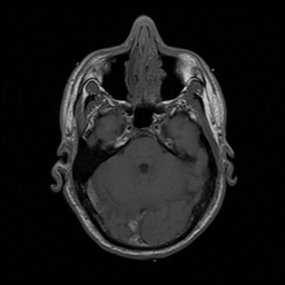
Anatomical contrast
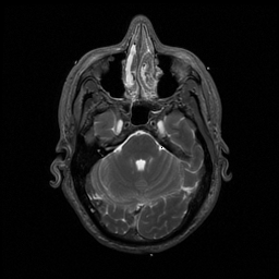
Different fluid and tissue contrast
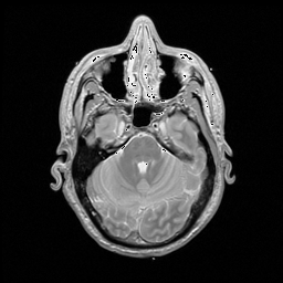
A further contrast setting
Courtesy of the U.S. National Library of Medicine · Visible Human male cadaver specimen · axial head sample m_vm1125 · retained 256 × 256 source-derived pixels · public-domain dataset. Display enlargement adds no acquisition detail. These retained historical samples do not represent the most current NLM dataset. This is cadaver research data, not a living-person clinical study or an NLM endorsement. Preparation and sequence conditions matter; no diagnosis or human medical review is claimed.
An axial image cuts across the head; a coronal view faces both hemispheres; a sagittal view separates left and right. Viewer left is not automatically the person’s left. The retained research images have no displayed patient-side marker, so this page does not infer laterality from them.
T1, T2 and proton-density images weight signal differently. Brightness is not a tissue’s natural colour. Sequence and specimen conditions influence appearance.
Structural MRI describes anatomy. MR angiography uses techniques that emphasize vessels and blood-related signal. BOLD functional MRI measures changes related to blood oxygenation; it is an indirect activity-related measure, not a photograph of thoughts.
Evidence: NIBIB / NIH
No authentic MRA, functional MRI, EEG trace or brain histology has been added. The atlas artwork, signal curve and sleep sequence are explanatory illustrations. They must not be read as acquired clinical evidence.
Read source and editorial limits →Evidence: Visible Human Project · NLM · MRI principles · NIBIB
Original English educational chapter. Scientific relationships checked against independent sources; generated illustrations are teaching aids, not factual evidence. Human medical review: Not performed. Physical-phone acceptance of this independent version remains Pending. The private book informed scope only; its prose and artwork are not reproduced.
New source pass: 9 October 2026. OpenStax anatomy, circulation, central processing, motor responses and NHLBI sleep material were accessible. NINDS brain and stroke material, MedlinePlus hydrocephalus and NCBI Circle of Willis were checked using available official indexed text; direct NINDS pages returned 403 and NCBI returned a browser challenge. These access limits do not establish new human medical review. This depth-completion pass additionally checked internal-capsule anatomy, published basal-nuclear and blood–brain-barrier reviews, memory resources, sleep regulation, cerebellar circuits and hypothalamic–pituitary interfaces. Relevant references also appear beside the new explanations. This chapter includes a superior surface view, choroid-plexus tissue interface, selected body sensory routes, separate venous/CSF outflow models and bounded hippocampal/plasticity mechanisms. New references are linked beside each lesson. Published reviews support the relationships; some NCBI/PMC direct pages returned browser challenges, so indexed primary-source excerpts were used and the access scope is recorded. Fine generated microstructure is illustrative. Source checks support the stated teaching relationships; they are not human medical review. No new authentic MRA, fMRI, EEG or histology is claimed. This final teaching pass pairs tissue and regional anatomy with the retained scientific mechanisms. Generated hippocampal folds are broad context, not a subfield atlas; sensory anatomy is not tractography; the SCN is located regionally rather than falsely drawn as a visible nucleus. The hippocampus reference remains an existing record; its current direct fetch was blocked. MRI credit and provenance retained from the existing chapter.