Similar colour does not mean identical layers
The gallbladder lacks a distinct muscularis mucosae and submucosa. It should not inherit the usual intestinal wall pattern simply because both organs contain a lumen.
GALLBLADDER & BILE ROUTES / THE 30-SECOND PICTURE
The liver makes bile. A muscular reservoir beneath it modifies and stores part of that supply. The cystic branch connects storage with a main route leading toward the duodenum.
Greater resistance at the intestinal outlet favors diversion through the cystic duct into the gallbladder, where bile is concentrated. The liver continues making bile.
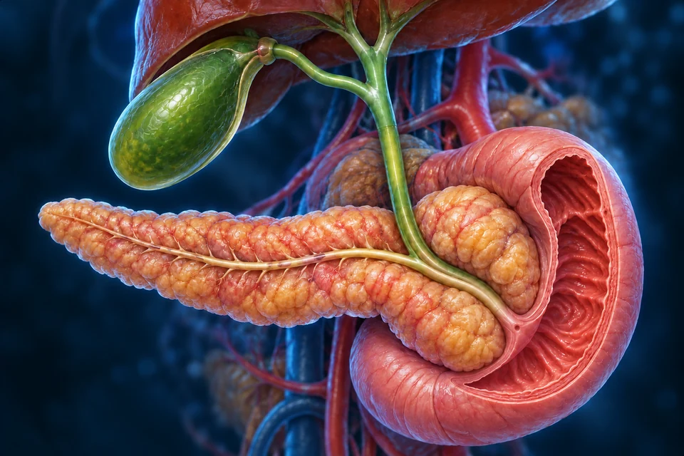
Gold shows local direction on the visible ducts. Liver bile production continues in both contexts; the illustration does not assign a fixed fraction to either route.
Structure and mechanism references: NIDDK · The normal biliary tract · Merck Manual Professional · Biliary function
01 / ORGAN → FOLD → EPITHELIAL SURFACE
Follow the broad fundus into the body and narrowing neck. The inner folds face bile. A closer window separates one epithelial layer, connective support and deeper interlacing muscle.
Follow the broad fundus through the body toward the neck. The folded lining faces the stored bile; muscle lies deeper in the wall.
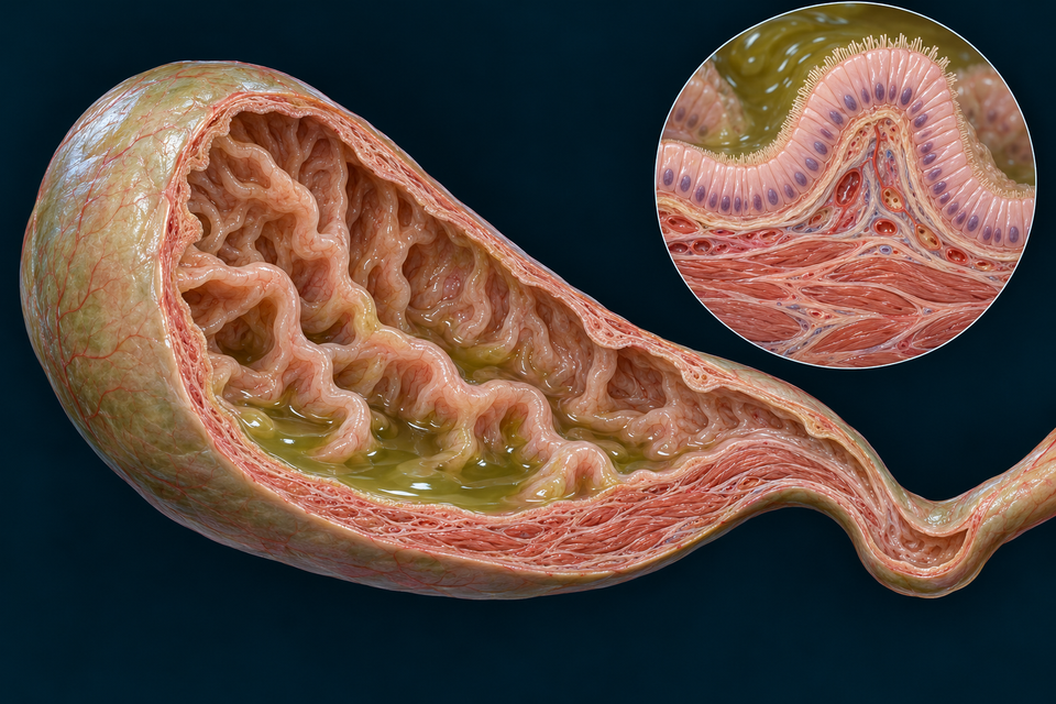
The gallbladder lacks a distinct muscularis mucosae and submucosa. It should not inherit the usual intestinal wall pattern simply because both organs contain a lumen.
A mucosal fold is a tissue shape. Microvilli are much smaller extensions on individual cells. Both can add surface, but they are not the same structure.
Connect the lining with epithelial organisation →Structure and mechanism references: Digital Histology · Gallbladder wall · SIU School of Medicine · Bile ducts and gallbladder
02 / LESS SOLVENT, A MORE CONCENTRATED RESERVOIR
Read from luminal bile across the epithelial sheet toward the tissue circulation. Select the compartment to connect the visible structure with its role in concentration.
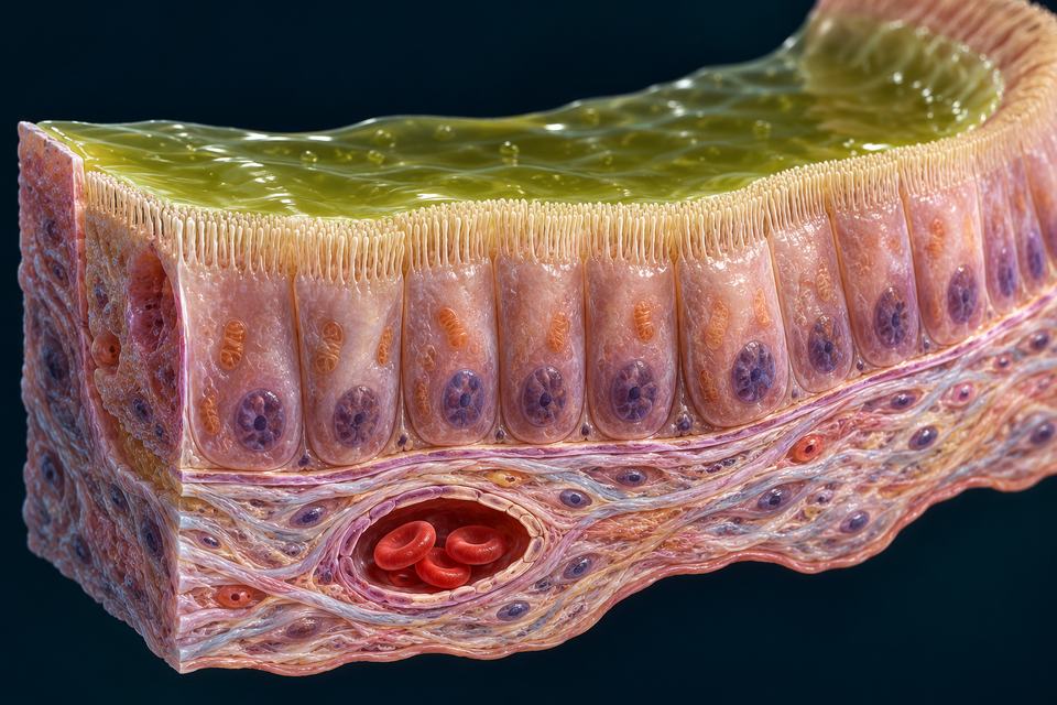
LUMEN → EPITHELIUM → TISSUE RETURN
The liver has supplied the bile. The luminal fluid contains water, electrolytes and organic constituents. Recovering part of the water changes concentration without creating a new batch of bile.
Structure and mechanism references: SIU School of Medicine · Bile ducts and gallbladder · OpenStax · Accessory digestive organs · Merck Manual Consumer · Gallbladder and biliary tract
03 / A MEAL REACHES THE INTESTINE; THE RESERVOIR RESPONDS
Signals from the small intestine help recruit the stored supply. CCK and neural activity coordinate gallbladder contraction with relaxation of the terminal biliary outlet.
This is a hormonal and neural connection, not a new duct carrying food or CCK into the reservoir.
Intestinal acid stimulates bicarbonate-rich ductal secretion. Adding ductal fluid and recovering water from stored bile are different epithelial tasks.
Compare the pancreatic meal response → · Follow hepatic bile formation and delivery →
The two contexts on the opening image locate storage and release on the same anatomy. They do not portray a fixed fasting seal, a universal emptying fraction or every neural input.
Structure and mechanism references: Merck Manual Professional · Biliary function · OpenStax · Accessory digestive organs
04 / SEPARATE APPROACHES → A SHORT TERMINAL REGION
The local cutaway exposes what the regional view cannot resolve: a descending bile duct, a pancreatic duct, their selected short terminal channel and a small opening into the duodenal lumen.
The distal common bile duct brings bile from the liver–gallbladder system. It reaches a short local terminal region in the duodenal wall in this selected arrangement.
Gold follows the bile route. Blue follows the pancreatic route. Both approach the selected intestinal opening.

They help lipid digestion products interact with the watery intestinal environment. They are not the enzyme lipase.
Continue with digestion and absorption →Enzymes and bicarbonate arrive through an exocrine route. Pancreatic hormones reach blood through a different route.
Compare pancreatic digestive products →Structure and mechanism references: Merck Manual Professional · Biliary function · NIDDK · The normal biliary tract
05 / INTESTINAL RECOVERY → HEPATIC REUSE → FEEDBACK
After helping in the intestinal lumen, much of the bile-acid pool returns through the living gut wall and portal circulation. Compare recovery with reservoir concentration, then connect return with hepatic transport and control of new synthesis.
Compare an intestinal recovery field with the qualified reservoir and liver preparations. They show three different organs and jobs, not consecutive frames of one cell or a single continuous duct.
The brush border faces intestinal contents. An absorptive cell spans the epithelial layer, while connective support and a separate blood vessel lie beneath it. The pale goblet cells supply a different epithelial function. This representative intestinal field is used to locate ileal transport; its appearance alone cannot establish the bowel segment.
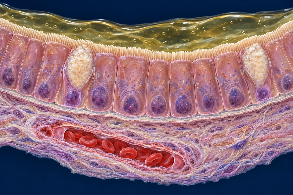
Gold rings locate the selected compartments. In the ileal material view, separate cyan arrows indicate apical entry and tissue-facing export; the gap preserves intracellular handling. They are not visible protein channels, flow measurements or a route through a broken epithelial layer. No arrow assigns blood-flow direction.
Mechanism evidence: Dawson et al. · Bile acid transporters · Ticho et al. · Intestinal absorption of bile acids · Zhang et al. · Human ileal explants and FGF19
Bile acids are made from cholesterol in the liver and are commonly conjugated with glycine or taurine before secretion. At intestinal pH much of this material is present as bile salts. Their amphipathic chemistry helps lipid products interact with the watery intestinal environment; pancreatic enzymes perform the chemical digestion.
Recovery occurs after luminal use. A mixed micelle does not enter an enterocyte as an intact chylomicron. Lipid products and bile acids can leave the luminal mixture by different mechanisms and ultimately take different routes. Follow the existing intestinal absorption lesson for lipid assembly and lymphatic entry.
Mechanism evidence: Ticho et al. · Intestinal absorption of bile acids · OpenStax · Liver, pancreas and gallbladder
The apical sodium-dependent bile acid transporter, ASBT (SLC10A2), is prominent in mature ileal absorptive cells. Its position at the brush-border membrane lets it take up bile acids from intestinal contents together with sodium. A transporter crosses a cell membrane; it is not a duct running through the whole tissue.
ASBT uses an existing electrochemical gradient. The Na⁺/K⁺-ATPase on the basolateral membrane consumes ATP to maintain low intracellular sodium. The energy-consuming pump and the sodium-coupled uptake carrier therefore occupy different functional steps. The drawing locates membrane domains, not verified individual protein structures.
Mechanism evidence: Dawson et al. · Bile acid transporters
After apical entry, bile acids are handled within the living enterocyte. Ileal bile-acid-binding protein contributes to intracellular handling; the cytoplasm is not an empty pipe. At the lateral and basal surfaces, the two-subunit OSTα/β system provides an important route of export.
OSTα/β is a facilitated transporter, rather than another sodium-driven ASBT or an ATP-burning pump. Transport direction depends on the prevailing conditions; net export is the normal absorptive context shown here. Export into the tissue compartment is followed by vascular exchange and downstream portal drainage.
Mechanism evidence: Dawson et al. · Bile acid transporters · Ticho et al. · Intestinal absorption of bile acids · Landrier et al. · FXR and human OST genes
The epithelial basal boundary, lamina propria and endothelial lining separate intestinal contents from blood. Absorbed bile acids traverse this tissue relationship before returning through intestinal veins toward the hepatic portal system. The small illustrated mucosal vessel is not itself the entire portal vein.
Many long-chain dietary fats instead enter lymph as chylomicrons after cellular reassembly. Bile-acid recovery should not be drawn into that lacteal route merely because bile assisted fat absorption. The retained liver and intestine chapters continue the regional blood and lymph pathways.
Mechanism evidence: Ticho et al. · Intestinal absorption of bile acids · OpenStax · Liver, pancreas and gallbladder
At the sinusoidal surface, uptake systems including sodium-dependent NTCP and organic anion transporting polypeptides (OATPs) bring returning bile acids into hepatocytes. Their substrate contributions differ. These names identify molecular transport mechanisms; a coloured patch in an atlas cell is not evidence that a particular transporter has been imaged.
Canalicular BSEP uses ATP-dependent export to secrete bile salts toward the small bile space between adjacent hepatocytes. This is a different surface from the blood-facing uptake side. Canalicular bile continues into the biliary tree; blood remains in the sinusoidal circulation.
Mechanism evidence: Dawson et al. · Bile acid transporters · Ticho et al. · Intestinal absorption of bile acids
Bile acids also carry information. Activation of the nuclear receptor FXR in ileal cells can promote human FGF19 production. The resulting endocrine signal travels through blood rather than through a new duct between intestine and liver. FXR signalling can also regulate transport-related genes, including OSTα and OSTβ.
Human ileal explants exposed to bile acids showed increased FGF19 transcripts and secreted protein. This supports a responsive human tissue mechanism; the experimental concentrations and incubation times are not a timetable or diagnostic threshold for an individual after a meal. FGF19 expression is not exclusive to the ileum in every tissue or context.
Mechanism evidence: Zhang et al. · Human ileal explants and FGF19 · Landrier et al. · FXR and human OST genes · Human tissue-derived organoids · enterohepatic signalling
At the liver, FGF19 interacts with FGFR4 and the co-receptor β-Klotho. Downstream signalling can suppress CYP7A1 expression, reducing a major entry step in the classical pathway of new bile-acid synthesis from cholesterol. Returning material and a returning hormonal message are related, but they are not the same cargo.
Hepatic FXR-related mechanisms and wider metabolic inputs also regulate the pool. A human tissue-derived organoid system has linked intestinal bile-acid stimulation and functional FGF19 secretion with a hepatic response. Such controlled models illuminate the connection without reproducing every feature of an intact human circulation.
Mechanism evidence: β-Klotho and endocrine FGF signalling · Song et al. · FGF19 signalling in human hepatocytes · Human tissue-derived organoids · enterohepatic signalling
Water and electrolyte recovery from gallbladder contents leaves a more concentrated stored solution. Ileal recovery retrieves bile acids from the intestinal lumen after delivery. Both use polarised epithelia, but the organ, material and physiological purpose differ.
The reservoir is a side branch, not an obligatory checkpoint for every drop of liver bile. Reused bile salts can enter the continuing biliary supply whether or not that portion is temporarily stored. Keep formation, storage, concentration, delivery and recycling as connected but distinct processes.
Mechanism evidence: OpenStax · Liver, pancreas and gallbladder · Ticho et al. · Intestinal absorption of bile acids
Nutrients in the intestine stimulate hormonal and neural responses that coordinate bile delivery. In the existing meal-response lesson, CCK participates in gallbladder contraction and terminal outlet relaxation. Secretin has a different role in bicarbonate-rich ductal secretion.
The FXR–FGF19 connection taught here concerns feedback on bile-acid synthesis. It should not replace the meal-related motor explanation. These are selected dominant teaching roles, not a claim that each hormone has only one effect or that all responses occur simultaneously.
Mechanism evidence: OpenStax · Liver, pancreas and gallbladder · Zhang et al. · Human ileal explants and FGF19 · β-Klotho and endocrine FGF signalling
Enterohepatic cycling conserves much of the available bile-acid pool, but recovery is not perfect. Some material escapes absorption and is lost in faeces. New hepatic synthesis replenishes this loss while signalling helps coordinate production with the returning pool.
The balance varies with bile-acid species, intestinal transit and physiological context. No fixed recovery percentage, daily cycle count or universal synthesis rate is assigned by these states. This lesson explains normal control, not a test of whether a person has malabsorption.
Mechanism evidence: Ticho et al. · Intestinal absorption of bile acids · Dawson et al. · Bile acid transporters
The intestine contains a mixture of bile-acid species. Microbial deconjugation and other transformations change this mixture, including formation of secondary bile acids. Chemical form affects membrane permeability and which transport mechanisms can contribute.
Passive absorption can occur outside the main active ileal route, particularly for suitable unconjugated species. Therefore “all bile acids return only through ASBT in the terminal ileum” is too absolute. The illustrated ASBT–OST path explains a major organised recovery mechanism without depicting every microbial or passive route.
Mechanism evidence: Ticho et al. · Intestinal absorption of bile acids
Human FGF19 and the related mouse FGF15 are not interchangeable labels for the same experiment. The linked human explant, human cell and organoid studies answer different questions; animal pathway experiments add evidence but cannot supply a universal human response size.
The new intestinal image and retained gallbladder and hepatocyte images are original generated teaching illustrations, not acquired microscopy. They support visible compartment relationships. Receptor conformation, transporter counts, enzyme location and precise ultrastructure have not been certified from their appearance. The functional claims come from the accompanying sources.
Mechanism evidence: Zhang et al. · Human ileal explants and FGF19 · Song et al. · FGF19 signalling in human hepatocytes · Human tissue-derived organoids · enterohepatic signalling · β-Klotho and endocrine FGF signalling
No. ASBT couples uptake to sodium movement. The basolateral Na⁺/K⁺-ATPase spends ATP to maintain the sodium gradient; these are different proteins and steps.
No. Apical uptake, intracellular handling and basolateral export precede tissue and vascular exchange. Blood stays in its endothelial compartment.
No. Reservoir concentration chiefly recovers water and electrolytes from stored bile; ileal recovery retrieves bile acids after intestinal use.
No. Feedback on new synthesis and transport of an existing molecule are different processes. Hepatic uptake and canalicular secretion remain part of reuse.
Normal physiology sources checked 21 October 2026. Human tissue studies, experimental models and generated atlas detail are distinguished above. No personal diagnostic or treatment thresholds are provided. AI-assisted source checks do not constitute physician review or fine-anatomy certification.
GO DEEPER / STRUCTURE, TRANSPORT, CONTROL AND CONNECTIONS
The gallbladder sits against the inferior surface of the liver in the right upper abdomen. A broad fundus continues into the body, which narrows into the neck and cystic duct. The organ is muscular, but it does not make the liver’s supply of bile.
Right and left hepatic ducts unite into the common hepatic duct. The cystic duct joins this main route; the channel below that junction is the common bile duct. Naming the segments makes it possible to follow a route without mistaking a side branch for the only way forward.
The adjacent pancreas and duodenum explain the close approach of two secretions near the intestinal wall. They do not make bile a pancreatic product. In an opened preparation, removal of covering tissue exposes relationships that would otherwise be hidden.
Structure and mechanism references: NIDDK · The normal biliary tract · OpenStax · Accessory digestive organs · Merck Manual Consumer · Gallbladder and biliary tract
The liver continues forming bile between meals. The relative resistance of the downstream outlet helps divert a portion into the gallbladder through the cystic duct, while some bile can continue toward the intestine.
When the reservoir empties, the cystic direction reverses: stored fluid returns to the main duct. One connecting duct can therefore serve filling and emptying at different times. It is not a permanently one-way pipeline.
Meal size, composition and timing influence delivery. A drawing with two learning contexts should not be read as a universal timetable, a completely sealed fasting outlet or a fixed proportion of bile entering storage.
Structure and mechanism references: Merck Manual Professional · Biliary function · Merck Manual Consumer · Gallbladder and biliary tract
A simple columnar epithelial sheet follows the mucosal folds. Supporting lamina propria forms their core and contains vessels and other tissue cells. Deeper irregular smooth-muscle bundles change the shape of the sac.
The normal gallbladder lacks a distinct muscularis mucosae and submucosa. Its smooth muscle is not separated into the neat inner circular and outer longitudinal coats used to describe much of the intestinal muscularis externa.
Outer covering also depends on location: connective tissue attaches the hepatic surface to the liver, whereas the free peritoneal surface has a serosal covering. A selected free-wall cutaway is not a map of the entire attachment.
Structure and mechanism references: Digital Histology · Gallbladder wall · SIU School of Medicine · Bile ducts and gallbladder
Broad branching folds allow the mucosa to occupy a changing internal shape and provide substantial epithelial surface. At cell scale, apical microvilli add finer exchange area. The fold and the microvillus are different levels of organisation.
The lining cells are tall and face the lumen on one side, supporting tissue on the other. Their polarity matters: exchange across a barrier requires distinct entry and exit surfaces. It cannot be explained by drawing green bile directly connected to a red blood vessel.
A gallbladder section can resemble intestine at first glance. The pattern of folds, lining and deeper wall must be read together; a pink projection alone does not establish a villus or an intestinal crypt.
Structure and mechanism references: SIU School of Medicine · Bile ducts and gallbladder · Digital Histology · Gallbladder wall
The concentrating role depends on net absorption of water and electrolytes across the mucosa. Less solvent remains in the reservoir, so many bile constituents become more concentrated. This is a change to bile already supplied by the liver, not new hepatic synthesis within the gallbladder.
Solute transport and water movement are coupled. Active solute handling helps establish local driving forces; water recovery is not performed by a fictional bucket-shaped pump. Channels, carriers and energy-dependent transport are different mechanisms, even when their combined effect is net fluid absorption.
Classic experiments on isolated rabbit and guinea-pig gallbladders helped investigate this coupling. Those experimental preparations do not supply a universal human rate or identify the proteins in a generated illustration. The learning image keeps the main barrier and direction clear without pretending to measure either.
Structure and mechanism references: OpenStax · Accessory digestive organs · SIU School of Medicine · Bile ducts and gallbladder · Diamond · 1964 · Experimental salt and water transport
Smooth-muscle activity changes reservoir geometry and can drive contents toward the cystic duct. Effective delivery also depends on the connected outlet: resistance downstream and activity in the reservoir must be coordinated.
Concentrating and emptying are different jobs. The mucosa exchanges material across the wall; contraction delivers the luminal contents along a duct. A cell-to-capillary arrow and a gallbladder-to-duodenum arrow therefore describe different routes.
The illustrations compare roles and anatomical positions. They do not show an individual ejection fraction or prove that every part of the wall shortens identically during a real meal.
Structure and mechanism references: Digital Histology · Gallbladder wall · Merck Manual Professional · Biliary function · OpenStax · Accessory digestive organs
Nutrient-related signals in the small intestine promote release of cholecystokinin, or CCK. This gut hormone helps link gallbladder contraction with relaxation of the biliary outlet. Cholinergic neural activity participates in the response as well.
The signal travels through the body; it is not food moving backward up the bile duct to press a switch. The gallbladder does not manufacture CCK. Neural and endocrine influences help match delivery to a meal rather than setting a permanent on/off state.
Secretin has a different emphasis: it supports bicarbonate-rich ductal secretion in response to intestinal acid. That is not the same task as the gallbladder mucosa concentrating stored fluid. The pancreas and liver lessons develop the complementary secretory roles.
Structure and mechanism references: Merck Manual Professional · Biliary function · OpenStax · Accessory digestive organs
In a common arrangement, the distal common bile duct and main pancreatic duct meet near the duodenal wall and reach the major papillary opening through a short shared terminal region. Other connections and openings occur; one selected preparation cannot represent every variant.
The sphincter of Oddi is a muscle complex associated with the terminal duct region. Its components are tissue within this anatomical setting, not a large loose ring hung between two floating pipes. The local window exposes continuity without claiming every muscle fibre is resolved.
Bile and pancreatic juice approach from their own origins. Sharing an outlet does not normally mean bile is meant to flow backward into pancreatic ducts, and it does not turn insulin into a duct-borne digestive product.
Structure and mechanism references: Merck Manual Professional · Biliary function · NIDDK · The normal biliary tract
Bile includes water, electrolytes, bile salts, phospholipids, cholesterol and pigments such as conjugated bilirubin. These constituents do not all have the same purpose or fate.
Bile salts help lipids interact with the watery intestinal environment and support absorption of lipid digestion products. They are not pancreatic lipase and do not themselves perform all the chemical cleavage of dietary fat.
The reservoir modifies and times delivery. Most of the detailed manufacture, ductal modification and disposal chemistry belongs to the connected liver and intestinal lessons; following those links gives the process its full organ context.
Structure and mechanism references: Merck Manual Professional · Biliary function · Merck Manual Consumer · Gallbladder and biliary tract
The cystic artery is the main named arterial supply. It commonly arises from the right hepatic artery, but its origin and course vary. Small branches supply the organ wall; the generic vessels in the generated cutaway are not an operative arterial map.
Small venous channels communicate with hepatic and portal venous pathways. Regional lymph may reach cystic and hepatic nodes, including routes through the liver’s drainage. The wall is therefore connected to circulation and lymphatic tissue without being an open continuation of the bile lumen.
Autonomic input reaches the gallbladder through the hepatic plexus. Its neural context complements hormonal coordination. Naming the plexus explains a connection; it does not certify the branching pattern of fine nerves not exposed in these illustrations.
Structure and mechanism references: Kenhub · Cystic artery · Kenhub · Gallbladder neurovascular context · Merck Manual Professional · Biliary function
After bile enters the intestine, much of the bile-salt pool is recovered, especially near the terminal ileum, and returned through portal blood for hepatic uptake and resecretion. Storage is one part of this larger circulation, not its starting source.
Pigment handling is a separate story. Bilirubin and its intestinal products do not follow exactly the same path or purpose as recycled bile salts. The existing liver and small-intestine lessons separate reuse from elimination.
Normal concentration also does not imply that a stone must form. Disease depends on more than one illustrated feature. This chapter explains normal structure and function; it does not diagnose pain, recommend a procedure or select treatment.
Structure and mechanism references: Merck Manual Professional · Biliary function · Merck Manual Consumer · Gallbladder and biliary tract · NIDDK · The normal biliary tract
The generated plates expose selected structures in three dimensions. The linked institutional histology resources show real tissue in a section. Neither type of image answers every question.
For a section, start with the lumen, folded epithelial edge and supporting tissue, then locate deeper muscle. Section plane and preparation affect the appearance. Do not infer a cell’s exact transport proteins or flow rate just from its colour or shape.
These external specimen references are links, not copied slide pixels. Original anatomical artwork and editable labels complement them; they are not presented as authentic acquired microscopy.
Structure and mechanism references: Digital Histology · Gallbladder wall · SIU School of Medicine · Bile ducts and gallbladder
REAL TISSUE / COMPLEMENTARY EXTERNAL REFERENCES
Start at the luminal edge, trace the folded lining, then look for connective support and deeper muscle. The external resources show actual section material; the illustrations on this page are original generated teaching artwork.
Specimen identity and preparation are those described by each source. No institutional slide pixels are reproduced here, and a section is not a complete three-dimensional organ.
No. The gallbladder is a side reservoir; a direct downstream bile route also exists.
No. The mucosa recovers water and electrolytes from bile made upstream. Formation, concentration and emptying are separate tasks.
No. It is an original generated teaching illustration. The external section references provide a different kind of tissue evidence.
No. They retain distinct origins and roles even when their ducts share a short terminal passage.
Normal structure and function checked against the references below on 20 October 2026. The existing book inventory guided topic coverage and teaching scale, without copying its text or figures. This chapter does not diagnose symptoms or select treatment.
Original generated illustrations and retained qualified regional artwork use independent editable text and geometry. AI-assisted source checks and Owner visual standards do not certify every fine generated structure or replace physician review. Physical-phone and independent post-publication review remain separate.