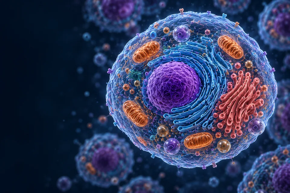
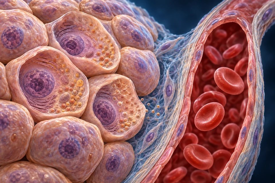
Air, nutrients, fluids and information cross boundaries between systems. Start with the cells that form tissues, then follow the organs, routes and responses that connect the body.
▷
Watch chapter overviewFilm planned · not yet available
Cell → tissue → organ → system · fine generated detail illustrative
02 / THORAX
Air and blood meet across a boundary.
Airway, lung and heart are distinct structures. A front-facing reference gives their spatial context; exchange happens at a much smaller scale.
30-SECOND ORIENTATION / THREE CONNECTED JOURNEYS
New original Golden-family organ preparation. Anterior orientation, anatomical right is image left. Only broad lung/heart/tracheal relationships are taught. Fine texture, fissure contours, vessels and hidden root connections illustrative / not independently verified. Not a patient image. Human medical review: Not performed.Structure notes · further context
Right lung
Broad visible organ context only. Small internal branches, exact surfaces and hidden vessel/airway roots are not independently verified by generated anatomy.
Left lung
Broad visible organ context only. Small internal branches, exact surfaces and hidden vessel/airway roots are not independently verified by generated anatomy.
Heart
Broad visible organ context only. Small internal branches, exact surfaces and hidden vessel/airway roots are not independently verified by generated anatomy.
Trachea
Broad visible organ context only. Small internal branches, exact surfaces and hidden vessel/airway roots are not independently verified by generated anatomy.
Air reaches lungs. Blood carries the exchanged gases.
The trachea branches toward lung air spaces. Pulmonary blood meets those spaces across a tissue boundary, then returns to the left heart. Air and blood do not become one tube.
Organs do not work in isolation. Start with their shared space, follow an exchange boundary, then explore the cells and signals that make the relationship work.
Anatomy
Where and what
Structure and location make a route understandable.
Physiology
How and why
Function asks what the structure does and how its activity is regulated.
Air moves through airways. Oxygen crosses from alveolar air toward pulmonary blood; carbon dioxide moves in the opposite exchange direction. The pathways connect functionally without becoming one tube.
ANATOMY AND RELATIONSHIP / ALL BASIC FACTS VISIBLE
Air, barrier and blood
Airway → alveolar air | exchange barrier | pulmonary blood
Cyan: oxygen toward blood · Gold: carbon dioxide toward air. These are qualitative exchange directions, not tracked particles or measured rates.
Original generated cellular atlas, not authentic microscopy. The supporting interval is expanded; it does not depict the thinnest fused interface at true scale. Fine membranes, organelles, nuclei and endothelial boundaries are illustrative. No macrophage is assigned to a non-resolved profile.
Air spaces
Ventilation brings air to and from the exchange region.
No blood tube is drawn inside the airway.
Exchange surface
Oxygen moves toward blood; carbon dioxide moves toward air.
The thin interface is a different scale from the source organ render.
Delivery
Blood carries oxygen onward through the left heart and systemic vessels.
Ventilation alone does not describe tissue delivery.
All essential relationships remain visible. Selecting a stage connects its explanation with anatomical context. The static preparations are not measured flow or a physiological simulation.
Why do lungs need both ventilation and circulation?
Ventilation renews gas in the alveolar spaces. Perfusion brings blood to the neighbouring exchange surface and carries it away again. Diffusion links the two sides across tissue; neither a blocked airway nor an unperfused air space is corrected merely by naming the other route.
After lung exchange, pulmonary veins return blood to the left heart. Systemic delivery then depends on pumping, blood carriage and the receiving tissue. The illustration separates gas space from the red-cell-containing blood compartment; it does not measure regional ventilation–perfusion matching.
The right heart sends blood toward the lungs. Pulmonary veins return it to the left heart, which sends it into the systemic circulation. Artery and vein describe direction relative to the heart, not a universal oxygen colour.
ANATOMY AND RELATIONSHIP / ALL BASIC FACTS VISIBLE
Follow the two linked circuits
Body → right heart → lungs → left heart → body
Generated organ context, not CT or a complete vessel-root reconstruction. Red/blue convention distinguishes relative oxygen content; hidden pulmonary veins and fine lung vessel trees are not individually mapped.
This anterior preparation locates heart, lung and proximal routes. Pulmonary veins and internal chambers are not all exposed; follow the chamber-level route in the linked Heart lesson rather than infer hidden joins from this surface.
Pulmonary arteries carry oxygen-poor blood in the normal adult route.
Return to left heart
Pulmonary venous return enters the left atrium.
A vein need not carry oxygen-poor blood.
Systemic circuit
Left ventricular output reaches body tissues and returns toward the right heart.
This is a route map, not exact vessel geometry.
All essential relationships remain visible. Selecting a stage connects its explanation with anatomical context. The static preparations are not measured flow or a physiological simulation.
Small-intestine regions sit near organs that process nutrients and supply digestive secretions. This expanded current anatomical preparation separates their broad teaching roles; it does not identify every hidden junction.
New original Golden-family text-free teaching preparation. Expanded regional orientation. Patient right is image left. Selected overlaps are opened for teaching, not a literal single-plane specimen. Hidden junctions, generated vascular detail, exact positions and proportions remain illustrative. Human medical review: Not performed.Structure notes · further context
Liver
A swallowed bolus does not travel through liver tissue. Bile exits by ducts; venous nutrient delivery is a different route.
Pancreas
The gland lies largely behind the stomach. Selective exposure reveals context, not normal unobstructed anterior visibility or exact duct openings.
Small intestine
Jejunum and ileum are not reliably separated by external color. Their regional and cell-scale differences belong to the linked deep lesson.
Colonic frame
The colon surrounds central loops in this expanded view. Real bowel positions vary; generated haustra are not a specimen count.
Digested material must cross the intestinal lining. Many absorbed nutrients reach the liver through blood; dietary lipid transport also involves intestinal lymphatics. Different nutrients do not all use one identical route.
ANATOMY AND RELATIONSHIP / ALL BASIC FACTS VISIBLE
Cross the lining before joining a delivery route
Intestinal lumen → epithelial transfer → blood or lymphatic transport
New Golden-family source-supported tissue relationships: lumen-facing villus, downward crypts and separate blood/lymph spaces. Individual generated cells, vascular branches and crypt details remain illustrative. The enlarged closed lacteal and vascular paths are selected teaching representations, not measured vessel diameters or an exact network.Structure notes · further context
Villus
Its core is supporting connective tissue, not an open digestive pipe.
Epithelial lining
Absorptive cells have apical microvilli. Junctions regulate passage between neighboring cells; the barrier is selective, not impermeable.
Lamina propria
It also contains immune and other supporting cells. A generated nucleus cannot establish a particular cell identity.
Blood vessels
The capillary network connects with larger vessels. Exact branch paths and red/blue vessel identities are illustrative.
Lacteal
Its tip is closed. It does not open into the lumen, and it does not carry every kind of absorbed nutrient.
Intestinal crypt
Crypts lie between villi. Stem-cell renewal and Paneth-cell roles are explained separately rather than guessed from generated cell colors.
Compare the epithelial boundary, blood network and central lymphatic context. The opened villus does not show each molecular transporter or assert that every lipid takes the same route.
Digestion changes material within the gastrointestinal lumen.
Being inside a gut lumen is not being inside a blood vessel.
Absorptive lining
Epithelial transport connects luminal material with tissue-side pathways.
Transporters and local barriers are explained in the Cells/Intestine lessons.
Distinct delivery
Many soluble nutrients enter portal blood; intestinal lipid transport also involves lymph.
This is not a universal single route for every molecule.
All essential relationships remain visible. Selecting a stage connects its explanation with anatomical context. The static preparations are not measured flow or a physiological simulation.
Why are digestion, absorption and delivery separate jobs?
Digestion changes material within the gastrointestinal lumen. Absorption transfers selected products across the lining into the body-side environment. Blood and lymph then provide onward delivery routes; calling the gut lumen “inside the body” does not make its contents part of the bloodstream.
Many water-soluble products reach the liver in portal blood. Much absorbed long-chain lipid leaves intestinal cells in particles that enter lymph before reaching venous blood. These routes differ by substance and processing; the regional organ picture is not a complete molecular transport map.
Kidneys regulate what remains in the blood and what leaves as urine. Ureters and bladder belong to the urine route; they are not renal blood vessels.
New anterior whole-tract context: person’s right is image left and the right kidney is slightly lower. Overlying organs are removed. Red/blue blood supply stays separate from ivory ureters and bladder. Fine vascular branches, adrenal texture, ureter insertion and generic urethral outlet are illustrative; no sex-specific continence anatomy is claimed.Structure notes · further context
Right kidney
In this anterior view it lies on image left. It is usually slightly lower than the left kidney because of the liver above it.
Left kidney
Image right is the person’s left. This view removes overlying organs to show the urinary structures, not their normal exposure.
Ureters
Peristaltic contractions propel urine; it does not depend only on gravity. Ureters are separate from renal vessels.
Bladder
The bladder wall relaxes during storage and contracts during coordinated emptying. Its depicted size is not a capacity measurement.
Urethra
Urethral length and surrounding anatomy differ between bodies. This generic outlet does not teach a male or female urethral course.
Why is filtrate not simply urine waiting to leave?
The glomerular filter separates a fluid pathway from blood. The tubule then changes that fluid by selective recovery and secretion. Water and useful solutes can return toward peritubular blood; other substances move toward the tubular lumen.
What finally reaches the renal pelvis and ureter is therefore different from the starting filtrate. Blood continues through the renal circulation and returns through renal veins. The paired ivory ureters in the organ view must not be mistaken for those blood vessels.
Intracellular fluid is inside cells. Extracellular fluid includes plasma within blood vessels and interstitial fluid around tissue cells. Other extracellular spaces include lymph and cerebrospinal fluid. These are not fixed-size tanks.
Exact current Endocrine text-free master reuse, with new independent compartment labels. Representative cell/blood preparation, not microscopy or measured fluid volumes. Fine membranes, granules and cell identities illustrative / not independently verified. Blood plasma is outside blood cells; their cytosol belongs to intracellular fluid. Human medical review: Not performed.Structure notes · further context
Cell interior
No amount, solute concentration or specific organelle identity is inferred from colour. Blood cells also have intracellular fluid.
Interstitial context
The fibres are support, not a direct visual measurement of fluid. Cell membranes and vascular walls are different boundaries.
Plasma context
The red cells are not plasma. The visible blood region provides compartment context, not a quantitative image of proteins, solutes or water.
SUPPORTING COMPARTMENT / RETURN MAP
Read the boundaries
Use the detailed cell–blood preparation directly above: the cell membrane encloses intracellular fluid, while the vascular wall separates plasma from the surrounding tissue. Match its three visible names to these boundaries.
The qualified anatomical preparations identify broad compartments and vessel-wall context. They do not measure fluid volumes, permeability or instantaneous flow.
Location changes the name
Intracellular fluid: enclosed within cell membranes. Blood cells also have intracellular fluid.
Interstitial fluid: outside tissue cells and outside blood vessels.
Plasma: the fluid component of blood, outside its cells.
Cell membranes and vascular walls are different boundaries. No fixed volume or automatic unrestricted transfer is encoded.
A capillary wall separates blood from interstitial space. A cell membrane separates the cell interior from its surroundings. Neither boundary is an unrestricted opening for every substance.
Vascular boundary
Plasma ↔ tissue interface
Permeability depends on the vessel bed and substance.
Cell membrane
Interstitial space ↔ cell
Channels, carriers and gradients matter.
Composition
Location changes the mixture
Plasma, interstitial and intracellular fluid are related, not identical.
Why are there two boundaries between plasma and a tissue-cell interior?
A vascular wall separates plasma from the tissue-side extracellular environment. A tissue-cell membrane then separates that interstitial environment from the cell interior. Water, ions, proteins and other molecules do not cross both interfaces by one unrestricted opening.
Interstitial composition reflects vascular exchange, cellular use and transport, and lymphatic removal. The endothelial surface layer matters, but a rat microvessel experiment is not a universal numerical flow rule for all human organs. The compartment illustration identifies locations without assigning fixed fluid volumes.
Lymphatic vessels collect interstitial fluid and proteins and return them toward venous blood. Do not read the tissue map as “all fluid exits at the arterial end and is absorbed at the venous end.”
SUPPORTING COMPARTMENT / RETURN MAP
Follow the return route
Original generated teaching preparation, not microscopy. Left-to-right is the reference direction for net transport. One nearly apposed valve pair is shown, not a complete pumping cycle or an entire lymphangion between serial valves. The controls locate different jobs; they do not animate leaflet opening.Structure notes · further context
A valve is not a motor
Pressure differences act on flexible leaflets. Opening permits forward passage; closure limits backflow. The valves themselves do not provide a second central heart.
Initial versus collecting vessel
Delicate initial lymphatics collect interstitial fluid through specialised endothelial arrangements. Collecting vessels have different wall organisation and intraluminal valves; this muscular preparation must not be relabelled as the blind-ended initial vessel above.
More than one source of propulsion
Intrinsic lymphatic-muscle activity and external tissue movement can both contribute. Which component dominates depends on the vessel and conditions; a fixed wave speed, contraction frequency or human flow rate is not inferred here.
This is a collecting vessel downstream of initial uptake. Its muscular wall and internal valve must not be relabelled as the delicate blind-ended initial lymphatic.
The qualified anatomical preparations identify broad compartments and vessel-wall context. They do not measure fluid volumes, permeability or instantaneous flow.
An additional return pathway
Tissue → initial lymphatic: collection begins from interstitial space.
Collectors → venous return: lymphatic vessels reconnect fluid and proteins with blood.
This is not a direct map of all branches. Do not infer routine complete reabsorption at every capillary’s venous end, or that no reabsorption ever occurs.
Actual exchange depends on the tissue and conditions; the route alone does not quantify flow.
A primary rat-microvessel experiment supports the importance of the endothelial surface barrier. It does not establish one flow rule for every human capillary bed.
Renal handling helps maintain water, salt and acid balance. The organ-level result depends on smaller-scale transport, not on a kidney acting as a simple waste sieve.
Neural pathways and hormonal signalling cooperate. A hormone can circulate widely, but a response depends on the receiving cell and its receptors. Neural versus hormonal does not mean “always fast” versus “always slow.”
ANATOMY AND RELATIONSHIP / ALL BASIC FACTS VISIBLE
A route and a receptor are different requirements
Signal production → delivery → responding target
New original Golden-family text-free teaching preparation. Focused lobe/stalk view adds tissue continuity rather than repeating the skull context. Anterior is image left. Solid endocrine tissue is distinct from neural texture; exact cell/vessel detail and scale remain illustrative. Human medical review: Not performed.Structure notes · further context
Hypothalamic floor
Specific neuron nuclei are not assigned from this generated appearance. Neural and vascular delivery require different mechanisms.
Infundibular stalk
The detailed portal connections are explained separately in an editable supporting map; the red/blue surface branches are not a verified angiogram.
Anterior endocrine tissue
The pars distalis forms the major endocrine region. Its cells are not exocrine acini around a drainage duct; generated cell subtype colors are not diagnostic.
Posterior neural tissue
The pars nervosa stores and releases hypothalamic vasopressin and oxytocin. It is not the site where those neuronal cell bodies synthesize their hormone precursors.
Vascular context
Exact arterial, long/short portal and venous junctions are not established by this generated surface. The functional route map states the source-supported main distinction.
A neuroendocrine example brings two routes together: hypothalamic axons end in posterior tissue; anterior secretory tissue has a different vascular connection. This is not a drawing of every neural synapse, receptor or target organ.
Activity travels through specialised neural pathways.
A synapse and a blood vessel are not interchangeable routes.
Hormonal route
A circulating hormone reaches many regions.
Receptor and cellular context influence which targets respond.
Local response
Cells change activity through the relevant signalling machinery.
Timing depends on the signal and response, not an absolute neural/hormonal speed rule.
All essential relationships remain visible. Selecting a stage connects its explanation with anatomical context. The static preparations are not measured flow or a physiological simulation.
Negative feedback reduces a disturbance to a regulated variable. A control diagram separates sensing, response and the resulting change; it is not a claim that every value stays perfectly constant.
ANATOMY AND RELATIONSHIP / ALL BASIC FACTS VISIBLE
Original generated medical illustration, not microscopy. The gland is enlarged for teaching; selected cut-open tubular profiles expose its lumen. These are not extra surface ducts. The view identifies broad compartments, not exact cell subtypes, channel proteins or measured layer thicknesses.Structure notes · further context
Follow a tube, not a branching tree
Eccrine glands are simple coiled tubular glands. The cut-open ends in this teaching view reveal selected parts of the coil; only one duct is followed to a surface opening.
Secretion and modification differ
Ion transport in the secretory segment supports fluid secretion. The duct recovers salt and has relatively low water permeability, so final sweat is usually less concentrated than plasma; composition varies with flow and adaptation. This plate does not depict individual transport proteins.
Cooling happens during evaporation
Sweat secretion moves fluid to the surface. Evaporation then removes heat; liquid that simply drips off does not provide the same evaporative cooling. Humidity, airflow and clothing affect evaporation.
Not the same as a kidney tubule
Sweat composition is not a direct reading of blood composition, and the duct is not drawn as a vasopressin-regulated water-recovery system. Kidney urine concentration and cutaneous cooling have different purposes.
Temperature regulation is one concrete example. Sensors and central integration lie beyond this gland window. Secretion reaches the surface through a duct; heat loss increases when that water evaporates. The arrow locates outward secretion, not a quantified heat flux.
A regulated variable provides information to the control process.
An organ picture alone does not show sensing.
Respond
Effectors change activity, transport or secretion.
Several tissues can participate in one response.
Feed back
The resulting change reduces the initial deviation.
Negative feedback is not the absence of variation.
All essential relationships remain visible. Selecting a stage connects its explanation with anatomical context. The static preparations are not measured flow or a physiological simulation.
What makes a response negative feedback?
“Negative” describes how the result acts on the original disturbance: the response tends to oppose it. It does not mean the response is harmful, that the variable never fluctuates, or that every regulated process uses one fixed central reference value.
In heat regulation, increased sweat delivery can support increased evaporation at the surface, while skin blood-flow changes alter heat transfer. A gland alone neither senses every aspect of body temperature nor guarantees cooling in every environment. The sensing, effector and resulting change are different parts of the explanation.
One signal may affect several tissues, and a single response may involve several signals. Follow the already detailed lessons rather than infer a complete mechanism from an organ’s proximity in a render.
Transport
Delivery and exchange
Heart, vessels and tissue interfaces connect the journey.
Control
Information and response
Neural, hormonal and local signals have different targets.
Response
Cellular work
Transport and tissue activity produce the organ-level change.
Anatomical models, imaging measurements and cellular preparations provide different kinds of evidence. A realistic-looking render is not a patient scan or a microscope observation.
ANATOMY AND RELATIONSHIP / ALL BASIC FACTS VISIBLE
Real scan and microscopy evidence has its own method, source and limitations. Visit the established chapter examples; this diagram does not manufacture an acquisition.
An interpretation must match the measured or constructed evidence. A single image can neither certify every hidden structure nor substitute for professional medical review.
A selected anatomical illustration or source-derived model explains spatial context.
Not a patient measurement or proof of hidden microscopic detail.
Acquisition
A physical signal is measured by a specific method.
Contrast, orientation and resolution depend on the acquisition.
Interpretation
A labelled educational explanation gives a bounded meaning.
A convincing visual appearance alone does not establish medical review.
All essential relationships remain visible. Selecting a stage connects its explanation with anatomical context. The static preparations are not measured flow or a physiological simulation.
Projection and section answer different questions.
A projection superimposes structures along a beam. CT reconstructs cross-sectional information from X-ray measurements. A rendered cut plane is a teaching operation, not a CT acquisition.
SUPPORTING GEOMETRIC COMPARISON / NOT MEDICAL IMAGING
Projection
Structures along the beam contribute to one projection. This original geometric map is not patient or simulated X-ray data.
Section
CT reconstructs sections from measurements. Selecting a cut plane in a teaching model is not a CT acquisition.
Why does an anatomical illustration not count as imaging evidence?
A projection combines information along paths through the body. CT reconstructs sections from many X-ray measurements. MRI, ultrasound, optical methods and nuclear imaging obtain different physical signals, with their own acquisition and reconstruction limits.
A beautiful cutaway may explain where structures lie, but its brightness and texture are designed rather than measured from a patient. The compact projection/section comparison represents geometry only. It must not be read as a synthetic scan, proof of a lesion, or guidance for choosing a clinical test.
Magnetic fields and radio-frequency pulses generate signals that can be reconstructed into images. The displayed contrast depends on acquisition; brightness is not a direct map of an organ’s natural colour.
A transducer sends sound and receives returning echoes. Position and acquisition affect the view. Doppler colours can encode motion relative to the probe; they must not automatically be read as oxygen-rich red and oxygen-poor blue.
Transducer
Sound emission and reception
Echo timing contributes spatial information.
Dynamic view
Frames over time
Movement can be observed without turning a still image into an anatomy master.
Colour key
Read the acquisition legend
A colour convention must be identified before assigning a meaning.
Endoscopy can show internal surfaces using optical instruments. Microscopy can reveal cells and tissue organisation; preparation, staining and optical method affect the resulting appearance.
Endoscopy
Internal surface view
A visible lumen surface does not expose every structure behind it.
Microscopy
Small-scale preparation
A section or field of view samples the tissue.
Rendered cells
Educational illustration
Illustrative texture is not a measured micrograph.
Nuclear imaging detects radiation associated with an administered tracer; the signal is interpreted through its distribution and protocol. An ECG records cardiac electrical activity. Neither is a direct photograph of every working cell.
PET / SPECT
Tracer-related measurement
PET detects annihilation photons; SPECT detects gamma emissions.
ECG
Electrical recording
A trace is not a photograph of valves opening.
Interpretation
Question and method together
Do not compare unlike outputs as if they were interchangeable.
The chapter teaches normal system relationships and method literacy. It does not diagnose disease, present simulated scan data as real, or replace the detailed organ lessons.
Represented
Distinct organ and tissue relationships
Three organ views and an expanded cell/blood interface; no repeated main anatomy image.
Not yet supplied
A comprehensive rights-cleared imaging gallery
Method comparison is present; new CT/MRI/ultrasound/PET specimen plates are not claimed.
Review
Source-supported stated scope
Human medical review and new physical-phone acceptance are not performed.
Selected normal system relationships, fluid compartments, feedback and imaging-method distinctions. Universal capillary-reabsorption cartoons, fixed fluid volumes and diagnostic claims are not adopted.
Methods and limits
AI-assisted original content and independent editable overlays. The qualified original thoracic preparation and exact current Cells, Digestive, Kidneys and Endocrine masters are retained. Qualified alveolar, cardiopulmonary, villus, pituitary, sweat-gland and collecting-lymphatic preparations connect regional context to mechanisms. Main anatomy teaches broad visible context; fine generated detail, hidden roots and individual variation remain illustrative / not independently verified. These images are not MRI, CT, microscopy or one person’s complete anatomical atlas. Historical HRA native geometry and CC BY 4.0 provenance remain preserved in source records; no old source files are deleted. Human medical review: not performed. Educational only.
Revision: integrated-body-normal-2026-10-19-eyes-v2. Stable local identity: ahb_ch_integrated_body_english_v1; not a new CMS entity. Text-free anatomical masters, semantic locations and translatable names stay separate; all16original plate targets are retained. Migration physical-phone acceptance: Pending, non-blocking. No clinical advice, physician approval or simulated medical images presented as measured evidence. HRA / HuBMAP United Female v1.10: CC BY 4.0; source attribution and transformation history are preserved.