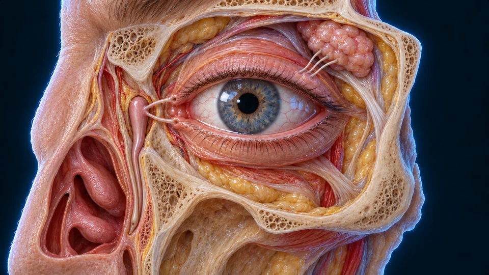
Explore the muscles that turn the eye, the iris that regulates light entry, and the lids, glands and fluid that maintain its exposed surface.
▷
Watch chapter overviewFilm planned · not yet available
Current Golden-family Hero · gross context · fine detail illustrative
Turn the globe. Adjust entry. Maintain a clear surface.
Three different systems support seeing: skeletal muscles direct gaze, iris smooth muscle controls the pupil, and lids with glands maintain the exposed surface. Choose a route to connect anatomy with its job.
30-SECOND ORIENTATION / TURN → ADMIT → MAINTAIN
Anterior is image left in both preparations. The corrected superior-rectus point follows the globe-facing belly, not the thinner band above it. Oblique deep insertion boundaries are not fully exposed.Structure notes · further context
Lateral rectus
The foreground straight muscle pulls the eye outward; motor supply is CN VI.
Superior rectus · visible belly
This upper straight muscle contributes elevation, intorsion and adduction; motor supply is CN III.
Superior oblique · trochlea
Its tendon turns through a superomedial pulley before returning toward the globe; motor supply is CN IV.
Superior oblique · reflected tendon
The returned tendon attaches behind the globe equator; it does not continue to the posterior muscle origin.
Inferior oblique · anterior route
The short curved muscle begins at the anterior medial floor and passes under the globe, unlike the recti. Its hidden full insertion is not mapped here; motor supply is CN III.
View 1 · Straight muscles on the globe
Anterior is left. Follow superior rectus toward its anterior scleral attachment. The thinner upper band remains unlabelled because its full endpoint is hidden.Structure notes · further context
Lateral rectus
The foreground straight muscle pulls the eye outward; motor supply is CN VI.
Superior rectus · visible belly
This upper straight muscle contributes elevation, intorsion and adduction; motor supply is CN III.
View 2 · Different oblique routes
The pulley turns the superior-oblique tendon back toward the globe. Hidden complete posterior insertions cannot be confirmed from this exposure.Structure notes · further context
Superior oblique · trochlea
Its tendon turns through a superomedial pulley before returning toward the globe; motor supply is CN IV.
Superior oblique · reflected tendon
The returned tendon attaches behind the globe equator; it does not continue to the posterior muscle origin.
Inferior oblique · anterior route
The short curved muscle begins at the anterior medial floor and passes under the globe, unlike the recti. Its hidden full insertion is not mapped here; motor supply is CN III.
Muscle pull turns the globe; a tendon can turn around a pulley.
Follow the globe-facing rectus muscle and the different oblique route. The neural supply coordinates actions; the eye does not move by six independent on/off arrows.
Four rectus and two oblique muscles turn the eye. The detailed orbital preparations distinguish globe-facing muscle bellies and the superior-oblique tendon turn. A seventh nearby muscle lifts the upper lid rather than turning the globe. The separate native left-eye reference can be opened below.
Anterior is image left in both preparations. The corrected superior-rectus point follows the globe-facing belly, not the thinner band above it. Oblique deep insertion boundaries are not fully exposed.Structure notes · further context
Lateral rectus
The foreground straight muscle pulls the eye outward; motor supply is CN VI.
Superior rectus · visible belly
This upper straight muscle contributes elevation, intorsion and adduction; motor supply is CN III.
Superior oblique · trochlea
Its tendon turns through a superomedial pulley before returning toward the globe; motor supply is CN IV.
Superior oblique · reflected tendon
The returned tendon attaches behind the globe equator; it does not continue to the posterior muscle origin.
Inferior oblique · anterior route
The short curved muscle begins at the anterior medial floor and passes under the globe, unlike the recti. Its hidden full insertion is not mapped here; motor supply is CN III.
View 1 · Straight muscles on the globe
Anterior is left. Follow superior rectus toward its anterior scleral attachment. The thinner upper band remains unlabelled because its full endpoint is hidden.Structure notes · further context
Lateral rectus
The foreground straight muscle pulls the eye outward; motor supply is CN VI.
Superior rectus · visible belly
This upper straight muscle contributes elevation, intorsion and adduction; motor supply is CN III.
View 2 · Different oblique routes
The pulley turns the superior-oblique tendon back toward the globe. Hidden complete posterior insertions cannot be confirmed from this exposure.Structure notes · further context
Superior oblique · trochlea
Its tendon turns through a superomedial pulley before returning toward the globe; motor supply is CN IV.
Superior oblique · reflected tendon
The returned tendon attaches behind the globe equator; it does not continue to the posterior muscle origin.
Inferior oblique · anterior route
The short curved muscle begins at the anterior medial floor and passes under the globe, unlike the recti. Its hidden full insertion is not mapped here; motor supply is CN III.
Compare the unchanged native source selection
Six muscles turn one globe
5
Left superior oblique
Its trochlear tendon route supports intorsion, depression and abduction.
More about left superior oblique
Fresh native projection and first named target surface raycast, transparent named target retained; not microscopic fibres,pump dynamics or clinical imaging. Broad selected roles independently source-supported; model detail is not human medical review.
4
Left medial rectus
Turns the globe toward the nose; oculomotor III supply.
More about left medial rectus
Fresh native projection and first named target surface raycast, transparent named target retained; not microscopic fibres,pump dynamics or clinical imaging. Broad selected roles independently source-supported; model detail is not human medical review.
2
Left inferior rectus
Depression with adduction and extorsion contributions.
More about left inferior rectus
Fresh native projection and first named target surface raycast, transparent named target retained; not microscopic fibres,pump dynamics or clinical imaging. Broad selected roles independently source-supported; model detail is not human medical review.
123456
6
Left superior rectus
Elevation with adduction and intorsion contributions.
More about left superior rectus
Fresh native projection and first named target surface raycast, transparent named target retained; not microscopic fibres,pump dynamics or clinical imaging. Broad selected roles independently source-supported; model detail is not human medical review.
3
Left lateral rectus
Turns the globe away from the nose; abducens VI supply.
More about left lateral rectus
Fresh native projection and first named target surface raycast, transparent named target retained; not microscopic fibres,pump dynamics or clinical imaging. Broad selected roles independently source-supported; model detail is not human medical review.
1
Left inferior oblique
An angled lower route contributes to extorsion, elevation and abduction.
More about left inferior oblique
Fresh native projection and first named target surface raycast, transparent named target retained; not microscopic fibres,pump dynamics or clinical imaging. Broad selected roles independently source-supported; model detail is not human medical review.
Six muscles turn one globe · names and basic functions are visible
Source-native geometry preserves selected component positions. Its coarse surface is a positioning reference, not the primary high-detail tissue preparation.
Adduction turns toward the nose; abduction turns away. Elevation and depression move the gaze up and down. Intorsion brings the top of the eye toward the nose; extorsion turns it away. These names refer to body directions, not simply left and right on a screen.
3-MINUTE MECHANISM / ANATOMY AND DISTINCT JOBS
Movements combine, rather than acting as single-axis switches
Anterior is image left in both preparations. The corrected superior-rectus point follows the globe-facing belly, not the thinner band above it. Oblique deep insertion boundaries are not fully exposed.Structure notes · further context
Lateral rectus
The foreground straight muscle pulls the eye outward; motor supply is CN VI.
Superior rectus · visible belly
This upper straight muscle contributes elevation, intorsion and adduction; motor supply is CN III.
Superior oblique · trochlea
Its tendon turns through a superomedial pulley before returning toward the globe; motor supply is CN IV.
Superior oblique · reflected tendon
The returned tendon attaches behind the globe equator; it does not continue to the posterior muscle origin.
Inferior oblique · anterior route
The short curved muscle begins at the anterior medial floor and passes under the globe, unlike the recti. Its hidden full insertion is not mapped here; motor supply is CN III.
View 1 · Straight muscles on the globe
Anterior is left. Follow superior rectus toward its anterior scleral attachment. The thinner upper band remains unlabelled because its full endpoint is hidden.Structure notes · further context
Lateral rectus
The foreground straight muscle pulls the eye outward; motor supply is CN VI.
Superior rectus · visible belly
This upper straight muscle contributes elevation, intorsion and adduction; motor supply is CN III.
View 2 · Different oblique routes
The pulley turns the superior-oblique tendon back toward the globe. Hidden complete posterior insertions cannot be confirmed from this exposure.Structure notes · further context
Superior oblique · trochlea
Its tendon turns through a superomedial pulley before returning toward the globe; motor supply is CN IV.
Superior oblique · reflected tendon
The returned tendon attaches behind the globe equator; it does not continue to the posterior muscle origin.
Inferior oblique · anterior route
The short curved muscle begins at the anterior medial floor and passes under the globe, unlike the recti. Its hidden full insertion is not mapped here; motor supply is CN III.
The same preparation stays visible in each state: elevation and depression include gaze-dependent combinations. The original model is not being moved as if one muscle had one exclusive action.
Horizontal
Adduction turns toward the nose; abduction turns away.
Vertical
Elevation and depression depend on coordinated muscle action and gaze position.
Torsion
Rotation around the visual axis accompanies other actions; this is not a diagnostic movement test.
Optional compact mechanism map
Qualitative supporting relationships; not measured muscle paths, pupil sizes or a universal blink-pump sequence.
Superior and inferior rectus contribute to vertical gaze and also to torsion and adduction. Oblique contributions depend on gaze position and include torsion. Do not assign each muscle one isolated arrow and assume that is its complete action.
Superior rectus
Above the globe
Elevation with intorsion and adduction contributions.
Inferior rectus
Below the globe
Depression with extorsion and adduction contributions.
Obliques
Angled routes
Superior: intorsion, depression and abduction. Inferior: extorsion, elevation and abduction.
Why can the same muscle help with more than one movement?
A muscle pulls along its anatomical route onto a globe that rotates within the orbit. The relation between that line of pull and the visual axis changes with gaze. Naming a primary contribution is useful, but it does not erase the other actions of the muscle.
For example, superior rectus contributes elevation, intorsion and adduction. Superior oblique follows a different course through the trochlea and contributes intorsion, depression and abduction. A pulley changing tendon direction is not an extra motor nerve or an attachment to the cornea.
The qualified illustration exposes selected visible bellies and tendon approaches. Hidden insertion borders are not thereby verified. It is a structural learning view, not a clinical eye-movement test or a complete surgical orbit.
The superior oblique passes through the trochlea in the upper nasal orbit before its tendon runs back toward the globe. The inferior oblique starts near the anterior orbital floor. Neither should be drawn as just another straight rectus.
Rectus muscles
Posterior origin
The four recti arise near the common tendinous ring.
Superior oblique
Trochlear turn
Its tendon route changes direction at a connective-tissue pulley.
Oculomotor nerve III supplies most globe movers; trochlear IV supplies superior oblique; abducens VI supplies lateral rectus. Levator upper-lid supply is also III. Eyelid closure uses facial VII, so it must not be placed in the six-muscle globe-movement group.
RELATIONSHIPS / DEFAULT-VISIBLE LEARNING
Globe movers and lid muscles have different nerve assignments
III: Supplies medial, superior and inferior rectus, inferior oblique and levator.
IV / VI: Trochlear IV supplies superior oblique; abducens VI supplies lateral rectus.
VII: Facial VII supplies orbicularis oculi for lid closure, not one of the six globe movers.
Optional compact relationship guide
Supporting categories, not histology or an exact neural tract/pump measurement. Fine anatomical context is taught in the main images.
Why distinguish turning the globe, lifting a lid and closing it?
The six globe movers rotate the eye. Levator palpebrae superioris raises the upper lid and is supplied by oculomotor III, but it is not a seventh globe mover. Orbicularis oculi closes the lids and is supplied by facial VII.
Among the globe movers, lateral rectus uses abducens VI and superior oblique uses trochlear IV. The other four use III. These assignments describe motor supply; they do not identify every sensory or autonomic fibre in the orbital region.
This distinction also separates protective blinking from a pupillary light response. A corneal sensory input and lid closure use V1 and VII; the parasympathetic pupillary output reaches the iris through III, the ciliary ganglion and short ciliary nerves.
Both eyes usually change gaze together toward a target, while near viewing also uses convergence. Muscle activity is coordinated through neural networks rather than one muscle working entirely alone. Vestibular stabilization and visual processing are taught in the linked chapters, not rebuilt as a generic six-arrow diagnosis test.
Paired gaze
Coordinated eyes
Conjugate movements generally direct both eyes in a common direction.
Convergence
Near target
Both eyes turn inward when needed for a near target.
Stabilization
Head movement
Vestibular and visual control help maintain useful gaze.
The iris is a tissue diaphragm around an opening: the pupil. A sector cutaway reveals the deeper circumferential sphincter and radial dilator arrangements. The optional native reference shows the separate coarse iris/cornea components; neither preparation is an acquired iris photograph.
The front stroma has been removed in one sector to expose deeper tissue. The radial pattern on the intact anterior surface is not itself a map of dilator muscle. This is an original teaching illustration, not a microscope image or measured iris thickness.Structure notes · further context
Read the cutaway
This selected exposure explains circular versus radial orientation and front-to-back relationships. It is not a measured cell count, isolated muscle specimen, clinical image or simulation of aperture size. Fine boundaries and attachments remain illustrative; no physician certification is claimed.
Compare the unchanged native source selection
The iris surrounds the light-entry opening
2
Left cornea
Translucent anterior context; the native fine surface is simplified.
More about left cornea
Fresh native projection and first named target surface raycast, transparent named target retained; not microscopic fibres,pump dynamics or clinical imaging. Broad selected roles independently source-supported; model detail is not human medical review.
12
1
Left iris
The tissue surrounds an opening; intraocular muscle fibres are not resolved.
More about left iris
Fresh native projection and first named target surface raycast, transparent named target retained; not microscopic fibres,pump dynamics or clinical imaging. Broad selected roles independently source-supported; model detail is not human medical review.
The iris surrounds the light-entry opening · names and basic functions are visible
Source-native geometry preserves selected component positions. Its coarse surface is a positioning reference, not the primary high-detail tissue preparation.
A circular sphincter constricts the pupil under parasympathetic control. Radially arranged dilator smooth muscle enlarges it under sympathetic control. These microscopic muscles are not separate meshes in the native iris plate; the supporting guide explains the relationship.
3-MINUTE MECHANISM / ANATOMY AND DISTINCT JOBS
Smooth muscle adjusts the light-entry opening
The front stroma has been removed in one sector to expose deeper tissue. The radial pattern on the intact anterior surface is not itself a map of dilator muscle. This is an original teaching illustration, not a microscope image or measured iris thickness.Structure notes · further context
Read the cutaway
This selected exposure explains circular versus radial orientation and front-to-back relationships. It is not a measured cell count, isolated muscle specimen, clinical image or simulation of aperture size. Fine boundaries and attachments remain illustrative; no physician certification is claimed.
Gold outlines locate the selected teaching region; the anatomical image remains unchanged. They do not measure force or depict a live change in pupil size. A smaller aperture and a rounder lens are different mechanisms.
Bright light
Parasympathetic sphincter activity narrows the opening.
Dim light
Sympathetic dilator activity helps enlarge the opening.
Qualitative supporting relationships; not measured muscle paths, pupil sizes or a universal blink-pump sequence.
Which tissue actually moves when a pupil changes size?
The iris is a thin living diaphragm in front of the lens. Its opening is the pupil. Circumferential sphincter muscle near the pupillary margin narrows that opening; radial dilator elements near the posterior surface contribute to enlargement.
The cutaway removes front stromal tissue in one sector so those different orientations can be seen. Visible radial surface texture on an intact iris should not be read as a photograph of its deeper dilator muscle. Colour and fibre thickness in the generated preparation do not establish cell identity or microscopic measurements.
Parasympathetic activity supports sphincter contraction, whereas sympathetic activity supports dilator contraction. The two targets have different neural routes. This lesson does not infer disease, prescribe a drug or ask the reader to test a pupil.
Retinal input travels with the optic pathway to pretectal midbrain regions. Bilateral connections reach parasympathetic output, then oculomotor III, ciliary ganglia and short ciliary nerves toward the iris sphincters. This broad reflex differs from the route supporting conscious visual interpretation.
RELATIONSHIPS / DEFAULT-VISIBLE LEARNING
Retinal input can influence both pupils
Input: Retinal light information reaches pretectal pathways; this is distinct from the conscious visual pathway.
Bilateral output: Pretectal connections influence parasympathetic nuclei on both sides.
Motor route: III, ciliary ganglion and short ciliary nerves link output to the iris sphincter. Sympathetic dilation is a different route.
Optional compact relationship guide
Supporting categories, not histology or an exact neural tract/pump measurement. Fine anatomical context is taught in the main images.
Lens accommodation changes optical power; convergence changes gaze direction; pupil constriction changes the aperture. They are distinct parts of the near response. The existing Eyes focusing lesson explains ciliary muscle and lens-support tension; no new lens deformation is inferred from these iris images.
Accommodation
Lens shape
The ciliary system changes focus.
Convergence
Eye direction
The globe movers turn both eyes toward a near target.
Why are accommodation, convergence and constriction not the same action?
Accommodation changes lens shape and optical power through the ciliary and zonular apparatus. Convergence rotates the two eyes toward a near target. Constriction reduces the pupil aperture through iris muscle.
These processes can cooperate during near viewing while acting on different structures. The iris sphincter should therefore not be substituted for the ciliary muscle, and a smaller pupil should not be described as the lens becoming rounder. Continue to the established Eyes focusing lesson for the optical comparison.
The lacrimal gland lies toward the upper outer side of the eye. Tears spread over the surface; drainage starts at the medial lid region and continues toward the nose. Selected source passages locate this relationship without drawing a nonexistent tube directly from gland to sac.
Original generated teaching context retained from Eyes & Vision. Nasal is image left. The gland has small delivery ducts toward the surface; it has no direct pipe into the lacrimal sac. Exact punctal apertures, valves and inferior nasal opening are not independently resolved.Structure notes · further context
Lacrimal gland region
A gland lies in the superior lateral orbit; ducts reach the conjunctival region.
Exposed eye and lids
Blinking spreads the tear film over the front surface.
Medial canalicular approach
Small channels from the lid margins lead toward the lacrimal sac.
Lacrimal sac context
The drainage sac lies near the medial orbital wall, beside the nose.
Nasolacrimal drainage approach
A descending duct carries drainage toward the nasal cavity.
Compare the unchanged native source selection
A gland supplies the surface; medial passages drain it
2
Left lacrimal canaliculus
Selected medial passage toward the sac; not all punctal detail shown.
More about left lacrimal canaliculus
Fresh native projection and first named target surface raycast, transparent named target retained; not microscopic fibres,pump dynamics or clinical imaging. Broad selected roles independently source-supported; model detail is not human medical review.
4
Left lacrimal sac
A medial passage receiving canalicular drainage.
More about left lacrimal sac
Fresh native projection and first named target surface raycast, transparent named target retained; not microscopic fibres,pump dynamics or clinical imaging. Broad selected roles independently source-supported; model detail is not human medical review.
5
Left nasolacrimal duct
Continues down toward the inferior nasal meatus.
More about left nasolacrimal duct
Fresh native projection and first named target surface raycast, transparent named target retained; not microscopic fibres,pump dynamics or clinical imaging. Broad selected roles independently source-supported; model detail is not human medical review.
Fresh native projection and first named target surface raycast, transparent named target retained; not microscopic fibres,pump dynamics or clinical imaging. Broad selected roles independently source-supported; model detail is not human medical review.
6
Left cornea
Translucent anterior context; the native fine surface is simplified.
More about left cornea
Fresh native projection and first named target surface raycast, transparent named target retained; not microscopic fibres,pump dynamics or clinical imaging. Broad selected roles independently source-supported; model detail is not human medical review.
3
Left lacrimal lake
Selected surface collection context near the medial lid region.
More about left lacrimal lake
Fresh native projection and first named target surface raycast, transparent named target retained; not microscopic fibres,pump dynamics or clinical imaging. Broad selected roles independently source-supported; model detail is not human medical review.
A gland supplies the surface; medial passages drain it · names and basic functions are visible
Source-native geometry preserves selected component positions. Its coarse surface is a positioning reference, not the primary high-detail tissue preparation.
Lacrimal glands contribute aqueous fluid; meibomian glands at the lid margins contribute lipids; ocular-surface mucins help maintain the wetted interface. The film is not three rigid independent sheets. A mucoaqueous region and surface lipid contribution interact during blinking.
Mucoaqueous region: Water, dissolved components and mucins form an interacting region with gradients.
Surface: Ocular-surface mucins help maintain wetting; schematic colour and thickness are not microscopy measurements.
Optional compact relationship guide
Supporting categories, not histology or an exact neural tract/pump measurement. Fine anatomical context is taught in the main images.
How can a film have different contributions without being three rigid sheets?
Lacrimal secretion contributes water and dissolved components. Meibomian glands at the lid margin contribute lipids, and mucins from the surface and secretory cells help maintain the wetted interface. These inputs are not identical to three separate solid laminae.
Blinking redistributes material across a dynamic surface. The lipid contribution, mucoaqueous region and surface-associated molecules interact; the functional film cannot be identified by three colours in a simplified drawing. The accompanying anatomical illustration locates supply and drainage rather than claiming to measure film thickness.
Orbicularis oculi helps close the lids; levator activity helps reopen the upper lid. Blinking spreads tear components and contributes to drainage. Exact lacrimal pump phases remain debated; this sequence shows the broad jobs, not a proven universal suction cycle.
3-MINUTE MECHANISM / ANATOMY AND DISTINCT JOBS
Closing and reopening support the exposed surface
Original generated teaching context retained from Eyes & Vision. Nasal is image left. The gland has small delivery ducts toward the surface; it has no direct pipe into the lacrimal sac. Exact punctal apertures, valves and inferior nasal opening are not independently resolved.Structure notes · further context
Lacrimal gland region
A gland lies in the superior lateral orbit; ducts reach the conjunctival region.
Exposed eye and lids
Blinking spreads the tear film over the front surface.
Medial canalicular approach
Small channels from the lid margins lead toward the lacrimal sac.
Lacrimal sac context
The drainage sac lies near the medial orbital wall, beside the nose.
Nasolacrimal drainage approach
A descending duct carries drainage toward the nasal cavity.
The image locates the open surface and drainage region; the three visible stages explain closure, reopening and redistribution. No new universal pump-phase sequence is inferred from the still image.
Close
Orbicularis oculi contributes to lid closure.
Reopen
Levator activity contributes to raising the upper lid.
Redistribute
Blinking spreads the film and assists tear transport; exact pump phases are not depicted.
Optional compact mechanism map
Qualitative supporting relationships; not measured muscle paths, pupil sizes or a universal blink-pump sequence.
Tears enter upper and lower puncta near the inner lid margins, pass through canaliculi toward the lacrimal sac, then down the nasolacrimal duct to the inferior nasal meatus. The native selection does not depict every valve, punctal opening or anatomical variation.
Puncta and canaliculi
Medial lid region
Small surface openings lead into the drainage passages.
Lacrimal sac
Medial collection route
Canalicular drainage approaches this larger passage.
Nasolacrimal duct
Toward the nose
The duct opens below the inferior nasal turbinate.
Why does fluid pass across an exposed surface before reaching the nose?
Small lacrimal delivery ducts open toward the superior conjunctival region. Fluid reaches the exposed ocular surface, is distributed with blinking, and approaches the medial lid margins. There is no direct gland-to-sac tube through the globe.
Upper and lower puncta lead to canaliculi, the lacrimal sac and then the nasolacrimal duct, which opens into the inferior nasal meatus. Tears are external surface fluid; aqueous humour belongs to a different internal compartment and circulation.
Blinking contributes to drainage, but detailed pump models do not all assign identical suction and compression phases. The still preparation and state controls preserve this uncertainty rather than making one universal hydraulic animation.
Spontaneous blinking, voluntary closure and protective reflexes are not identical mechanisms. In the corneal blink reflex, ophthalmic trigeminal V1 carries sensory input and facial VII activates closure muscles. This educational route is not an instruction to touch or test your cornea.
Spontaneous
Regular surface support
Blinking maintains the exposed interface.
Voluntary
Conscious closure
Higher control can close the lids.
Protective reflex
Sensory-triggered closure
V1 input and VII motor output contribute to corneal protection.
Extraocular globe movers and main eyelid movers are specialized skeletal muscle. Iris sphincter and dilator are smooth muscle. Gland tissue, conjunctival cells and the fluid film add other levels of support. These tissues cannot be inferred from the colour of a simplified 3D mesh.
Globe movement
Skeletal muscle
Coordinated motor control changes gaze.
Aperture control
Smooth muscle
Autonomic activity adjusts pupil size.
Surface support
Secretory tissue and fluid
Gland/surface contributions maintain a dynamic film.
The optional native-source plates locate selected structures; the original illustrations provide separate anatomical teaching context. Neither establishes fine iris fibres, actual tear-film thickness, gland acini, pupil-response speed or individual insertions. No clinical image or muscle test is presented as evidence.
Self-review found that the source globe context could obscure the iris opening with a pale surface. The anterior iris view now omits that context and uses only unchanged iris/cornea components. Orbital and lacrimal views omit iris detail and retain translucent globe context for location. The original geometry and first render attempts remain outsideGit. Fine source facets and surface seams remain limitations.
Light detection is only part of seeing. Neural control moves the eyes, iris muscles regulate entry, and lids/glands/fluid maintain the exposed surface. These additions complement frozen Eyes rather than replacing its approved structure, focusing, retinal or visual-pathway plates.
Selected eye movement anatomy, iris feedback, tear supply/drainage and blink support. Supporting mechanisms are not patient measurements.
Methods and limits
Original detailed Eyes orbital and lacrimal illustrations are reused with their qualified annotations; the reference chapter is unchanged. One new text-free iris cutaway exposes circular and radial tissue arrangements, with independent semantic names shared by the body, detail and accessibility layers. Original BodyParts3D positioning selections remain available as optional native references, not fine tissue primaries. Six accurate supporting guides are compact and optional; their facts remain visible. No native model deformation or new live3D, clinical ocular imaging, patient-specific measurement or generated microscopy claim. Human medical review: not performed.
Revision: ocular-support-eyes-context-2026-10-19-v1. Stable local identity: ahb_ch_ocular_support_english_v1; not a new CMS entity. Original masters stay outside Git; labels and paths remain editable. New physical-phone acceptance: not performed. No patient diagnosis, surgical instruction, medication advice or physician approval claim. Observed microscopy and clinical disease remain separate work.