Special Senses / VISUAL EXPLAINER
Where Does Fluid Inside the Front of Your Eye Drain?
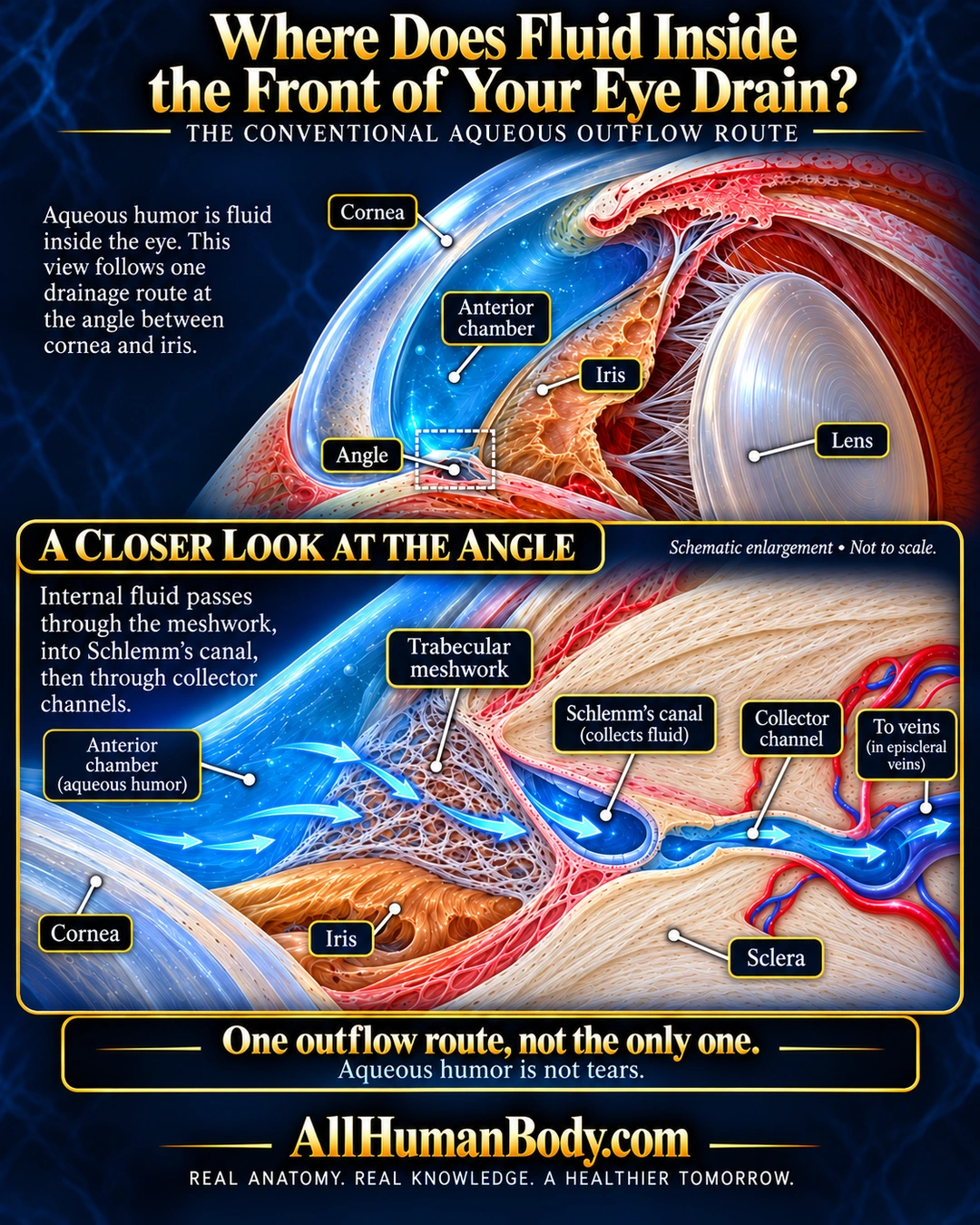
One route for fluid inside the front of the eye passes through the trabecular meshwork into Schlemm’s canal and collector channels. This aqueous-humor route is different from surface tear drainage and is not the only outflow route.
READ THE IMAGE
What the illustration shows
Navy and gold anatomy poster showing a sectional front of an eye, with a blue anterior chamber behind the cornea, peach iris and pale lens. A boxed angle is enlarged below. Blue arrows cross the trabecular meshwork, enter a blue Schlemm’s canal lumen and continue through a collector channel toward veins in ivory fibrous sclera. Labels identify the structures. Text specifies this is one outflow route, not the only one, and aqueous humor is not tears. Gold AllHumanBody.com branding and the full slogan appear below.
How to interpret this poster
- Illustrative channels and arrows are not measured flow or pressure. No glaucoma-causation, diagnosis or treatment claim; historical model assumptions excluded.
- AI-assisted educational illustration; not physician certification, microscopy, a diagnosis or individualized medical advice.
- Fine annotations need enlargement; the full description provides an accessible text alternative.
- Display the full uncropped illustration; do not trim titles, legends or boundary annotations.
AI-assisted educational illustration, adapted into web-sized images without changing its composition. This is general education, not an individual diagnosis or treatment recommendation.
EVIDENCE / CONTEXT
Sources and what they support
- Original aqueous-outflow modelling paper, university-hosted; page 2 flow description ↗
Trabecular meshwork → inner canal wall → Schlemm canal → collectors. Use stable anatomy only, not its historical glaucoma-causation hypothesis or numerical model.