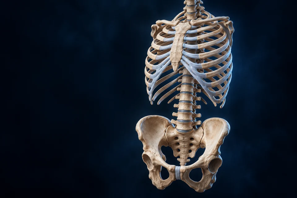
The chest protects vital organs while changing shape for breathing. The pelvic ring transfers load between trunk and limbs. Explore their bones, muscle partition and connective interfaces at different scales.
▷
Watch chapter overviewFilm planned · not yet available
Regional framework preparation · fine generated detail illustrative
02 / CAGE
The chest wall has both hard and flexible components.
Sternum lies anteriorly, the thoracic column posteriorly, and ribs curve between them. Costal cartilage contributes to the anterior framework.
Original Golden-family text-free teaching preparation. Anterior regional context, subject right image left. Broad sternum, rib and anterior cartilage relationships are source-supported. Conventional numbered attachment groups are explained separately; exact generated segment counts, cartilage boundaries, pores and individual shape remain illustrative / not independently verified. Human medical review: Not performed.Structure notes · further context
Manubrium
Clavicles meet lateral notches in life; broad clavicular context appears above the manubrium in this preparation. Fine articular borders remain illustrative. The first costal cartilage relates to the upper sternum.
Sternal body
The manubrium–body junction provides sternal-angle context. It is not the posterior vertebral column seen deeper.
Xiphoid process
Do not infer an age from the generated mineral texture. Soft tissues attach around this region.
Bony rib
Posterior joints and anterior cartilage relationships differ; the whole rib is not cartilage.
Costal cartilage
Costal cartilage is hyaline tissue; it is distinct from bone and from an interchondral connection.
Free anterior rib end
Floating ribs still attach posteriorly. Numbered classes are taught separately rather than counted from generated pixels.
The sternum lies at the front of a three-dimensional cage
Find the broad manubrium above the long body and smaller xiphoid. Ribs curve backward toward the thoracic column; the chest is an enclosure with openings, not a solid bony box.
Gold selects teaching regions; cyan arrows indicate a qualitative movement direction. Neither measures a force, flow, joint axis or respiratory phase.
The chest combines protection with changing respiratory volume. The pelvis connects trunk and limbs while providing bony and soft-tissue support. Neither is just a rigid box.
Thorax
Chest framework
Ribs, cartilage and sternum connect to the thoracic column.
Breathing also requires muscles and neural control.
Pelvis
Trunk–limb interface
Paired hip bones join the sacrum posteriorly and one another anteriorly.
Pelvic floor is soft tissue, not the bony ring.
Living interfaces
Bone, cartilage and connective tissue
Material differences make protection and motion compatible.
Three named sternal regions provide useful orientation.
Manubrium, body and xiphoid differ in shape and attachment context. The second costal cartilage relates to the manubrium–body junction; individual shape and ossification vary.
Manubrium
Upper broad segment
Clavicular and first-rib cartilage relationships orient the upper chest.
Not a thoracic vertebra.
Body
Elongated central segment
Anterior rib-cartilage relationships continue below the sternal angle.
A visible ridge does not define every joint.
Xiphoid
Small terminal region
Muscle/connective attachments surround a variable end.
The head, neck, tubercle and curved shaft serve distinct relationships. Typical middle-rib anatomy is not a template for every atypical rib.
Original Golden-family text-free teaching preparation. One representative typical middle rib, not a precise numbered specimen or complete joint. Head/neck/tubercle/shaft/anterior-end context is source-supported. Fine generated facet crests, groove, pores and individual curvature remain illustrative / not independently verified. Human medical review: Not performed.Structure notes · further context
Rib head
A typical middle rib head meets adjacent bodies near their interface. First, tenth, eleventh and twelfth ribs have exceptions; not every rib meets two vertebrae.
Rib neck
It is a continuous part of one bone, not a second rib branching from the shaft.
Rib tubercle
Costotransverse articulation is different from the rib-head contact; ribs eleven and twelve lack this articulation.
Rib shaft
A costal groove on the inner inferior border relates to vessels and nerve in life. Exact groove and neurovascular routes are not mapped here.
Anterior end
Cartilage is omitted to isolate bone shape. Atypical lower ribs do not have the same anterior attachment pattern.
Head and tubercle relate to different posterior contacts
In a typical middle rib, the head meets vertebral-body facets and the tubercle meets a transverse-process facet. The neck lies between these regions; atypical ribs have important exceptions.
Gold selects teaching regions; cyan arrows indicate a qualitative movement direction. Neither measures a force, flow, joint axis or respiratory phase.
A typical rib head relates to vertebral bodies; its tubercle meets a transverse process. The head is not a forked bone attached independently to two separate shafts.
Costovertebral
Head / body-region interface
A typical middle-rib head relates to adjacent vertebral-body facets.
First and lower-rib patterns differ.
Costotransverse
Tubercle / transverse process
A separate contact connects the rib near its neck.
Ribs11and12 lack this articulation.
Segmental movement
Joint + ligament + cartilage context
Motion is distributed across connected interfaces.
No fixed angle follows from the close-up.
Why do head and tubercle contacts teach different connections?
In a typical middle rib, the head and tubercle connect with different vertebral surfaces. Those posterior contacts, anterior cartilage connections and surrounding muscles together influence movement. Atypical first and lower ribs differ, so one isolated middle rib cannot establish every level or joint.
Cartilage connections have different names and materials.
Rib–cartilage, cartilage–sternum and cartilage–cartilage interfaces are not interchangeable. The first sternocostal connection differs from most later ones.
Costochondral
Rib → its cartilage
A primary cartilaginous interface links different tissues.
Sternocostal
Cartilage → sternum
First and later connection types are not uniformly identical.
Supporting relationship map, not exact anatomical geometry or a measured time course. All steps are visible before highlighting.
Original Golden-family text-free teaching preparation. Anterior regional context, subject right image left. Broad sternum, rib and anterior cartilage relationships are source-supported. Conventional numbered attachment groups are explained separately; exact generated segment counts, cartilage boundaries, pores and individual shape remain illustrative / not independently verified. Human medical review: Not performed.Structure notes · further context
Manubrium
Clavicles meet lateral notches in life; broad clavicular context appears above the manubrium in this preparation. Fine articular borders remain illustrative. The first costal cartilage relates to the upper sternum.
Sternal body
The manubrium–body junction provides sternal-angle context. It is not the posterior vertebral column seen deeper.
Xiphoid process
Do not infer an age from the generated mineral texture. Soft tissues attach around this region.
Bony rib
Posterior joints and anterior cartilage relationships differ; the whole rib is not cartilage.
Costal cartilage
Costal cartilage is hyaline tissue; it is distinct from bone and from an interchondral connection.
Free anterior rib end
Floating ribs still attach posteriorly. Numbered classes are taught separately rather than counted from generated pixels.
Bone meets its own costal cartilage
Find the transition from bony rib to pale anterior cartilage. A primary cartilaginous connection here must not be confused with a joint between cartilage and sternum or between neighbouring cartilages.
Gold selects teaching regions; cyan arrows indicate a qualitative movement direction. Neither measures a force, flow, joint axis or respiratory phase.
The diaphragm is a living partition, not a sealed wall.
A muscular dome and central tendon separate chest from abdominal context. Openings and posterior passage relationships remain necessary; the teaching window omits organs.
Original Golden-family text-free teaching preparation. Representative resting anterior cutaway; subject right image left. Domed muscle/tendon and chest-versus-abdominal relationships are source-supported. Fine generated fibres, crura, openings, attachment detail and exact dome height remain illustrative / not independently verified. This static view is not a measured respiratory phase. Human medical review: Not performed.Structure notes · further context
Diaphragm muscle
Contraction changes shape and displaces the partition inferiorly during inspiration. This is not a solid barrier without passages.
Central tendon
It is connective tissue rather than another chamber or a patch of bone.
Lower costal context
Sternal, costal and lumbar attachments participate. Exact tiny insertion sites are not independently verified in generated fibres.
Thoracic space
Heart and lungs are deliberately omitted, not absent in life. Pleural and pericardial compartments require their own teaching views.
Abdominal context
The abdominal cavity is not identical to the chest or a rigid sealed container. Pressure relationships vary with activity.
Peripheral muscle converges toward a central tendon
The domed partition combines contractile muscle with a non-contractile tendinous centre. Its peripheral attachment and posterior relationships connect it with the lower thoracic framework.
Gold selects teaching regions; cyan arrows indicate a qualitative movement direction. Neither measures a force, flow, joint axis or respiratory phase.
Movement changes cavity dimensions before air enters.
Diaphragm contraction moves the partition inferiorly; rib movement changes front-to-back and side-to-side dimensions. The lungs follow chest mechanics through their surrounding relationships.
Vertical
Diaphragm contraction → inferior displacement
Chest vertical dimension increases without requiring a perfectly flat dome.
Front–back
Pump-handle context → sternal/anterior motion
Rib movement contributes to the anterior–posterior dimension.
Side–side
Bucket-handle context → lateral rib motion
Movement contributes to transverse expansion.
Supporting relationship map, not exact anatomical geometry or a measured time course. All steps are visible before highlighting.
Original Golden-family text-free teaching preparation. Representative resting anterior cutaway; subject right image left. Domed muscle/tendon and chest-versus-abdominal relationships are source-supported. Fine generated fibres, crura, openings, attachment detail and exact dome height remain illustrative / not independently verified. This static view is not a measured respiratory phase. Human medical review: Not performed.Structure notes · further context
Diaphragm muscle
Contraction changes shape and displaces the partition inferiorly during inspiration. This is not a solid barrier without passages.
Central tendon
It is connective tissue rather than another chamber or a patch of bone.
Lower costal context
Sternal, costal and lumbar attachments participate. Exact tiny insertion sites are not independently verified in generated fibres.
Thoracic space
Heart and lungs are deliberately omitted, not absent in life. Pleural and pericardial compartments require their own teaching views.
Abdominal context
The abdominal cavity is not identical to the chest or a rigid sealed container. Pressure relationships vary with activity.
A descending diaphragm increases the vertical dimension
Muscle contraction generally moves the diaphragm inferiorly. The arrows are a qualitative direction on an anatomical still, not measured deformation, airflow through the muscle or a universally flat dome.
Gold selects teaching regions; cyan arrows indicate a qualitative movement direction. Neither measures a force, flow, joint axis or respiratory phase.
Supporting dimensional comparison, not a simulation of measured lung volume. No perfectly flat dome or universal displacement is asserted.
How does a moving chest wall lead to airflow?
Inspiratory muscle action increases thoracic dimensions. Through the pleural relationship, lung volume increases and alveolar pressure can fall below atmospheric pressure, drawing air inward. This is separate from gas exchange across an air–blood interface. During quiet expiration, elastic recoil contributes to outward airflow; forced breathing recruits additional muscle activity.
Muscle action depends on location and breathing demand.
External intercostals and parasternal/intercartilaginous internal parts can assist inspiration. Interosseous internal parts contribute to forced expiration. Quiet expiration commonly relies on elastic recoil.
Between ribs
Regional muscle layers
Fibre orientation and location affect mechanical action.
Not every internal part has one expiratory role.
Quiet breathing
Diaphragm and coordinated chest motion
Recoil helps the relaxed return.
Not obligatory forceful abdominal contraction.
Increased demand
Additional muscle recruitment
Neural and muscular contributions vary with activity.
Breathing links a structural wall to nervous regulation.
Normal movement requires coordinated respiratory neural output and muscle activation. Local rib shape alone cannot explain rhythm, gas exchange or voluntary breath control.
Posterior and anterior connections form one load-sharing ring.
The two hip bones connect through sacrum and anterior pubic union. Their lateral cups relate to the femoral heads; the pelvis is not simply one enlarged vertebra.
Original Golden-family anterior pelvic-ring teaching preparation, with a separately corrected normal four-pair anterior sacral foramen pattern. Paired hip bones, posterior sacral and anterior pubic connections are broad source-supported teaching relationships. Fine generated pores, cartilage, attachments and individual proportions remain illustrative / not independently verified. Not a sex classifier, birth predictor or operative atlas. Human medical review: Not performed.Structure notes · further context
Iliac wing
Its inner fossa and upper crest differ from the lateral acetabulum. Adult ilium is continuous with ischium and pubis.
Sacrum
It is separate from each hip bone. Fine foraminal routes and segment counts are not a surgical map.
Sacroiliac joint context
Cartilage-bearing and ligamentous regions coexist; the two bones are not universally fused into one adult bone.
Pubic symphysis
This is not the hip socket or a freely mobile ball-and-socket joint.
Acetabulum
The head and its joint tissues are omitted; a cup alone is not the whole hip joint.
Obturator foramen
In life a membrane largely spans this opening; it is not an unobstructed passage for a whole pelvic organ.
The sacrum connects the column with paired hip bones
Sacroiliac joint regions lie on either side of the sacrum. Cartilage-bearing surfaces and ligamentous support contribute different parts of the connection; normal support does not mean that all these bones are fused.
Gold selects teaching regions; cyan arrows indicate a qualitative movement direction. Neither measures a force, flow, joint axis or respiratory phase.
Three developmental regions become one adult hip bone.
Ilium, ischium and pubis converge around the acetabulum. Adult continuity does not erase their different surfaces, attachments and roles.
Original Golden-family text-free teaching preparation. Lateral regional view, anterior image right. Broad ilium/ischium/pubis/cup/opening/notch context is source-supported. Fine generated gluteal lines, cartilage, labrum, pores and individual proportions remain illustrative / not independently verified. No exact muscle attachment or complete socket joint is claimed. Human medical review: Not performed.Structure notes · further context
Iliac crest / wing
Attachment and support surfaces continue below it; crest shape varies among individuals.
Acetabulum
The articular rim and central nonarticular region have different roles. Detailed cartilage and labrum are not resolved here.
Pubic region
On the full ring its medial face participates in the pubic symphysis; this isolated bone does not show the opposite half.
Ischial tuberosity
Soft tissue distributes contact in life. This is not a pressure measurement or a posture prescription.
Obturator opening
An obturator membrane and associated muscle tissues are omitted for bony clarity.
Greater sciatic notch
Ligaments transform regional notches into passages in life. This plate is not a detailed nerve-route map.
The ilium forms the broad upper part of the hip bone
This is a lateral right hip-bone preparation. The iliac crest borders the superior wing; the view is not the medial iliac fossa. Real contours and proportions vary between individuals.
Gold selects teaching regions; cyan arrows indicate a qualitative movement direction. Neither measures a force, flow, joint axis or respiratory phase.
An inlet, an outlet and a floor describe different things.
The brim outlines entry to the lesser pelvis. The outlet is its inferior boundary, partly completed by ligaments. A musculofascial floor supports contents while permitting passages.
Inlet
Superior boundary of lesser pelvis
Bony landmarks define a transition from the greater pelvic context.
Not a solid lid.
Outlet
Inferior boundary
Bony and ligamentous margins define a lower opening.
Not the obturator foramen.
Floor
Living soft-tissue support
Muscles/fascia support pelvic viscera and functional passages.
Not a sealed bony plate.
Why is a visible bony opening not the pelvic floor?
Inlet and outlet describe boundaries of a regional space. The obturator opening is a separate bony feature, largely closed by membrane in life. Muscle, fascia and connective support contribute to a living floor beyond these bone surfaces. This bony preparation cannot identify every hidden soft-tissue boundary or predict birth capacity.
Femoral-head contact, cartilage, labrum, capsule and surrounding muscles contribute to the full articulation. The bony plate intentionally isolates the acetabulum.
Hip mobility and sacroiliac load transfer have different jobs. The anterior symphysis is another material interface, not a third hip socket.
No new femoral geometry, canonical female reconstruction or operative attachment map is introduced.
Stable support does not mean universal bony fusion.
Sacroiliac regions include cartilage-bearing joint context and strong ligamentous connections. Their structure and mobility change with individual development and age.
Articular context
Anterior/inferior regional surfaces
Opposing surfaces permit limited relative motion.
Generated visible borders do not resolve every cartilage type.
Ligamentous context
Posterior/superior support
Strong connective structures restrain separation and motion.
Not an empty space or universally fused bone.
Whole-ring context
Spine + pelvis + active support
Load transfer also depends on muscles and surrounding connective tissues.
The pubic symphysis joins paired medial surfaces through an interpubic disc and surrounding support. Small physiological motion differs from a freely moving synovial joint.
Original Golden-family text-free teaching preparation. Local connective-interface teaching window, not a measured histology slide or exact normal joint width. Pubic bone/disc/cartilage/ligament context is source-supported. Fine generated fibres, pores, clefts, vessels and layer thickness remain illustrative / not independently verified. No universal microscopic arrangement or synovial ball socket is asserted. Human medical review: Not performed.Structure notes · further context
Pubic bone section
Generated pores represent broad bone context, not measured marrow or individual trabeculae.
Interpubic disc
It resists different loading directions while allowing limited movement; it is not an intervertebral nucleus or a hip cavity.
Articular-covering context
Hyaline covering and fibrous disc are distinct. Exact thickness cannot be read from this generated plate.
Superior fibrous support
Fine fibre attachments are illustrative; the literature does not resolve every universal attachment.
Pubic ramus
The local cutaway isolates an interface, not the entire surrounding pelvis or floor.
Paired pubic surfaces face a connective interface
Sectioned bone lies on each side. Articular coverings and the intervening disc are different material regions; the enlarged preparation does not measure cartilage thickness or represent acquired histology.
Gold selects teaching regions; cyan arrows indicate a qualitative movement direction. Neither measures a force, flow, joint axis or respiratory phase.
Why does a small-motion union differ from a hip socket?
The pubic symphysis uses an intervening fibrous-cartilaginous disc and supporting tissues. The mobile hip is a synovial articulation with a femoral head, acetabular cartilage, labrum and capsule. These different interfaces belong to one connected region but do not share identical material structure or movement.
A material comparison explains why the joints behave differently.
Mineralised bone, cartilage, fibrous disc and ligament do not share one microscopic organisation. Cells maintain matrices that distribute forces and exchange nutrients.
Chondrocyte context
Cartilaginous matrices
Maintains extracellular matrix; tissue region changes phenotype.
No universal cell portrait from generated fibres.
Fibrous-disc context
Interpubic union
Collagen-rich tissue resists several loading directions.
Fine cleft and fibre geometry remain uncertain.
Ligament context
Around pelvic joints
Fibrous support constrains relative motion.
Exact attachments vary and are not a surgical map.
Standing and sitting change where support is carried. The sacrum, hip bones, joints, ligaments and muscles interact, rather than one bone working alone.
Standing
Column ↔ sacrum / SI ↔ hip bones / hips
Lower-limb support and trunk forces interact through the ring.
Sitting
Pelvic ring ↔ ischial / soft-tissue contact
Support distribution changes with posture and surrounding tissues.
Active restraint
Muscle + ligament + joint surfaces
Stability is shared, not supplied by one passive block.
Supporting relationship map, not exact anatomical geometry or a measured time course. All steps are visible before highlighting.
Original Golden-family anterior pelvic-ring teaching preparation, with a separately corrected normal four-pair anterior sacral foramen pattern. Paired hip bones, posterior sacral and anterior pubic connections are broad source-supported teaching relationships. Fine generated pores, cartilage, attachments and individual proportions remain illustrative / not independently verified. Not a sex classifier, birth predictor or operative atlas. Human medical review: Not performed.Structure notes · further context
Iliac wing
Its inner fossa and upper crest differ from the lateral acetabulum. Adult ilium is continuous with ischium and pubis.
Sacrum
It is separate from each hip bone. Fine foraminal routes and segment counts are not a surgical map.
Sacroiliac joint context
Cartilage-bearing and ligamentous regions coexist; the two bones are not universally fused into one adult bone.
Pubic symphysis
This is not the hip socket or a freely mobile ball-and-socket joint.
Acetabulum
The head and its joint tissues are omitted; a cup alone is not the whole hip joint.
Obturator foramen
In life a membrane largely spans this opening; it is not an unobstructed passage for a whole pelvic organ.
The ring connects axial and lower-limb support
Trunk and lower-limb forces interact through sacral, sacroiliac, hip-bone and hip-joint relationships. Gold selects these regions without claiming one fixed one-way path, force magnitude or load percentage.
Gold selects teaching regions; cyan arrows indicate a qualitative movement direction. Neither measures a force, flow, joint axis or respiratory phase.
Supporting relationship map, not a quantitative model or exercise prescription. Forces can travel in both directions through the framework.
Levator-ani and coccygeus context, fascia and connective attachments contribute to visceral support. Openings and coordinated function remain essential.
This regional bone chapter does not claim a full pelvic-floor muscle atlas. Reproductive, urinary and digestive relationships require their own source-verified views; detailed named fascial boundaries remain staged.
Hip-bone regions grow around developing cartilage before fusion. Sternal and cartilage ossification, joint surfaces and pelvic proportions vary through life.
Developing framework
Growth and changing interfaces
Early separate regions and cartilage contribute to adult form.
No single birthday for complete fusion.
Maturing tissues
Matrix and surface changes
Ossification does not progress identically in all structures.
No age classification from one image.
Individual form
Shape / sex-related averages / variation
Averages do not define every person.
No childbirth prediction or binary sex classifier.
Thoracic structure relates to lungs and heart; pelvic structure to limbs, urinary, digestive and reproductive support. Bone also participates in wider matrix and mineral maintenance.
A realistic illustration and an observation answer different questions.
An illustration selects a useful view. Imaging captures defined signals from a real subject; microscopy samples tissue at a different scale. Neither can be substituted by generated fine texture.
Gross illustration
Recognisable regions and interfaces
Supports first-glance location and function.
Not patient anatomy or exact count measurement.
Imaging / microscopy
Real signals or sampled tissue
Future independently licensed references can add evidence.
No new patient pixels acquired or fabricated.
Mechanism comparison
Selected normal relationships
Explains movement, material and load context.
Not a measured diagnostic or therapeutic simulation.
This chapter connects chest and pelvic foundations to preceding skull/column and subsequent limb teaching. It does not claim a complete respiratory, pelvic-floor or neurovascular atlas.
Clinical disease comparison, diagnosis and treatment remain deferred. Fine generated foramina, fibres, cartilage thickness and microscopic detail remain illustrative / not independently verified. Human medical review has not been performed.
No duplicated primary view, public draft release, database publication change or full-system completeness percentage.
Broad chest/rib/diaphragm/pelvic/hip-bone/symphysis relationships; selected normal material, movement, load, growth and cross-system functions. Authoritative references are used critically rather than repeating every simplified or historical claim.
Explicit limits
AI-assisted visual/content production. Fine generated vascular, foraminal, matrix, cartilage, muscle and attachment details remain illustrative / not independently verified. Human medical review: not performed. Educational only; not diagnostic, clinical or surgical material.
Revision: thorax-pelvis-english-2026-10-02-deep-v1. Stable local key: ahb_ch_thorax_pelvis_english_v1. No invented canonical CMS/atlas entity or 3D dataset. Seven original Golden-family masters are retained; a separately versioned pelvic-ring correction clarifies the normal four-pair anterior sacral foramen pattern. Thirty-three semantic targets keep editable names and explanations separate from anatomy. Native teaching contexts and source-linked deeper readings extend the normal foundation. Fine generated ribs/foramina/fibres/matrix and exact articular or muscle attachment topology are illustrative / not independently verified. No precise individual count, measured respiratory phase or birth prediction is inferred. Migration physical-phone acceptance: Pending, non-blocking.
The retained source-guided Vertebral Column card derives from BodyParts3D (Database Center for Life Science, CC BY-SA 2.1 Japan) and Z-Anatomy (CC BY-SA 4.0; PC export by Lluís Vinent Juanico), with AllHumanBody camera, materials, lighting and AI-assisted refinement. That card retains CC BY-SA 4.0; no source geometry or mixed-license datasets are supplied here. Original independent thoracic and pelvic illustrations are not acquired patient images.