Special Senses / VISUAL EXPLAINER
What Makes the White of the Eye a Tough Outer Coat?
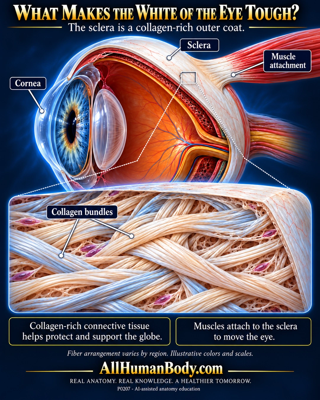
The sclera forms most of the tough outer coat of the eye. Its collagen-rich connective tissue helps support the globe and provides attachment for muscles that move it.
READ THE IMAGE
What the illustration shows
A navy-and-gold anatomy poster titled What Makes the White of the Eye Tough? An eye cutaway has a transparent anterior cornea at left, ivory sclera around the globe and a coral muscle tendon attaching externally. Labels point to the cornea, sclera and muscle attachment. A connected enlargement below shows interlacing ivory collagen bundles with scattered purple cells. Two boxes explain structural support and muscle attachment. Footer carries AllHumanBody.com and the full slogan. Colors and dimensions are illustrative.
How to interpret this poster
- Fiber arrangement varies by region; wall thickness, vessel pattern and enlarged collagen are illustrative rather than measured microstructure.
- The muscle is an attachment example, not a named-muscle insertion map. Deeper ocular layers are context, not complete histology.
- No four-layer classification, trauma guidance, eye-color diagnosis or evolutionary explanation.
- AI-assisted educational illustration; not physician certification, microscopy, a diagnosis or individualized medical advice.
- Fine annotations need enlargement; the full description provides an accessible text alternative.
- Display the full uncropped illustration; do not trim titles, legends or boundary annotations.
AI-assisted educational illustration, adapted into web-sized images without changing its composition. This is general education, not an individual diagnosis or treatment recommendation.
EVIDENCE / CONTEXT
Sources and what they support
- Cleveland Clinic: Sclera ↗
Collagen-rich outer coat, structural support and extraocular muscle attachment.
- Characterization of collagen from normal human sclera ↗
Human scleral collagen composition; biochemical study abstract, not fiber-geometry measurement.
Evidence correspondence is limited to the abstract.