Musculoskeletal / VISUAL EXPLAINER
How Tendons Bend Your Fingers
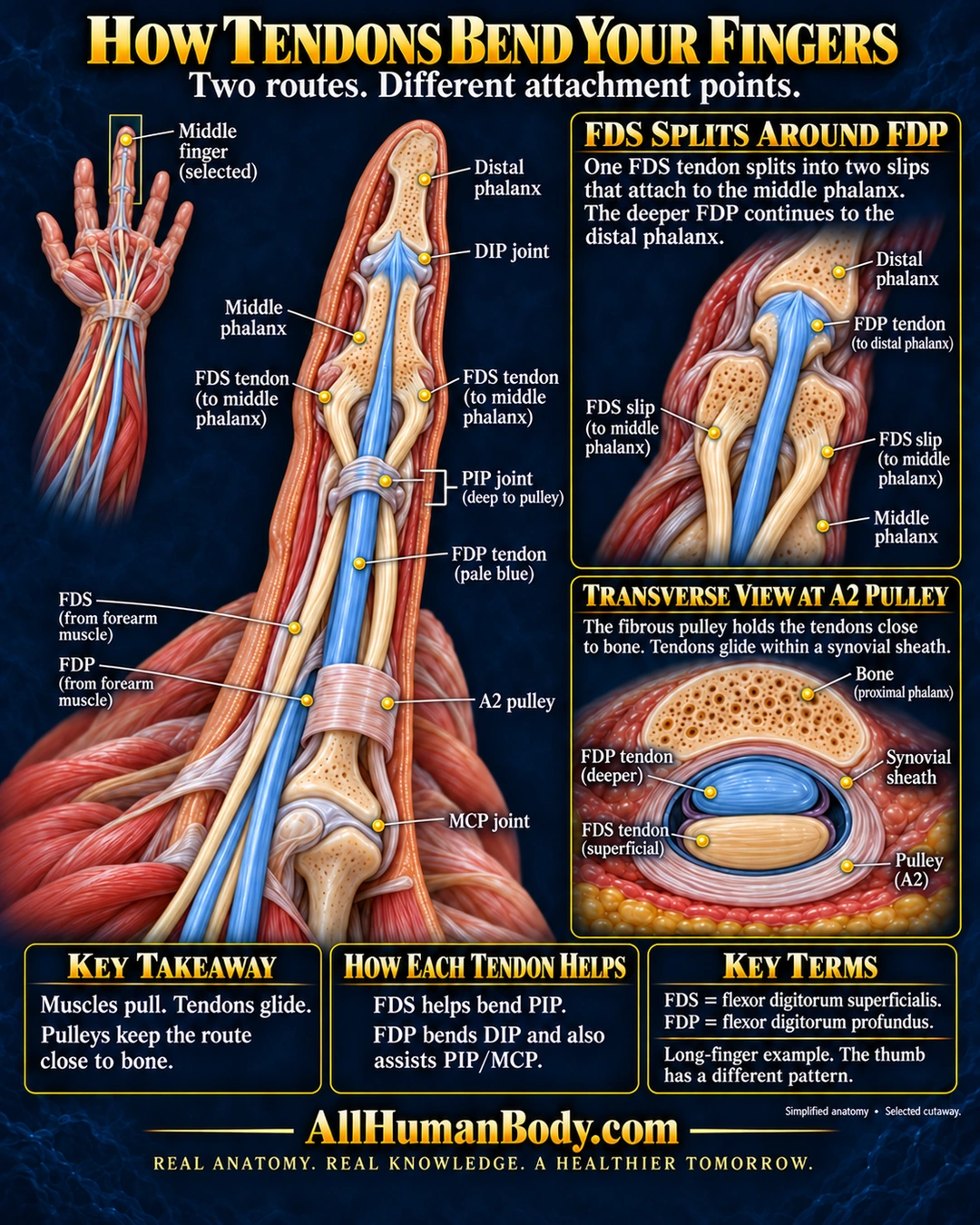
In the four longer fingers, the superficial flexor tendon divides around the deeper tendon. Their different attachment points help explain how they bend different joints while pulleys keep the gliding tendons close to bone.
READ THE IMAGE
What the illustration shows
Anatomical infographic titled “How Tendons Bend Your Fingers: Two routes. Different attachment points.” A small palm-side view highlights the middle finger. A large selected cutaway shows cream flexor digitorum superficialis (FDS) splitting into two slips that attach to the middle phalanx, while pale-blue flexor digitorum profundus (FDP) passes between them and attaches to the palmar base of the distal phalanx. DIP and MCP joint levels and an A2 pulley are labeled; a bracket marks the PIP joint level with the clarification “deep to pulley.” An enlarged inset shows the FDS split around FDP. A transverse A2 inset places FDP nearer the proximal phalanx and FDS more superficial, with synovial sheath and a fibrous retaining pulley around the tendons. Takeaways explain that muscles pull, tendons glide, pulleys keep tendons close to bone, FDS helps bend PIP, and FDP bends DIP and assists PIP/MCP. The thumb has a different pattern. Gold AllHumanBody.com branding and the slogan “REAL ANATOMY. REAL KNOWLEDGE. A HEALTHIER TOMORROW.” appear below.
How to interpret this poster
- The middle finger is a representative example; the thumb has a different arrangement.
- Selected coverings are removed; this is not a complete pulley inventory or a surgical map.
- The PIP joint is deep to the depicted pulley, not fully exposed in the main view.
- AI-assisted educational illustration; not physician certification, microscopy, a diagnosis or individualized medical advice.
- Fine annotations need enlargement; the full description provides an accessible text alternative.
AI-assisted educational illustration, adapted into web-sized images without changing its composition. This is general education, not an individual diagnosis or treatment recommendation.
EVIDENCE / CONTEXT
Sources and what they support
- Tendons ↗
FDS/FDP routes, attachments, muscle-driven tendon action and thumb distinction.
- Flexor tendon mechanism ↗
Long-finger flexor routes, joint contributions, sheath and pulley relationships.
- Anatomy of the flexor tendon sheath and pulley system ↗
Organization of the synovial-lined fibro-osseous tendon tunnel.
Evidence correspondence is limited to the abstract.