Musculoskeletal / VISUAL EXPLAINER
Can a Foot Have an Accessory Navicular Bone?
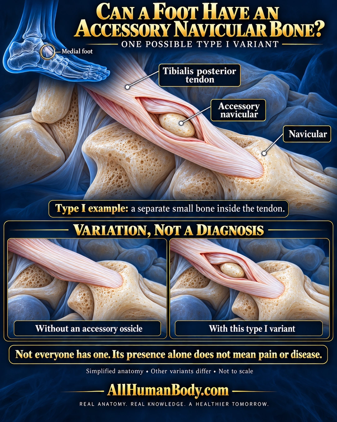
A small accessory navicular can occur near the native navicular on the inner foot. This poster shows one type I example enclosed within the distal tibialis posterior tendon; its presence alone does not establish pain or disease.
READ THE IMAGE
What the illustration shows
Navy-and-gold anatomy poster titled “Can a Foot Have an Accessory Navicular Bone?” A small blue medial-foot locator circles the inner midfoot. The enlarged central illustration shows a pale pink, fiber-textured tibialis posterior tendon passing diagonally toward a larger ivory navicular bone. An illustrative cutaway window reveals a small oval accessory navicular enclosed by tendon fibers, separate from the native bone. Direct labels identify tendon, accessory navicular and navicular. Two lower matched illustrations compare a tendon without an accessory ossicle with the pictured type I variant. Text states that not everyone has one and its presence alone does not mean pain or disease. A note says simplified anatomy, other variants differ and not to scale. Gold AllHumanBody.com branding and the full slogan sit below.
How to interpret this poster
- Single cadaver evidence supports a possible tendon relationship, not prevalence or all accessory forms. Window is an illustrative exposure, not a tendon tear; no diagnostic or treatment inference.
- AI-assisted educational illustration; not physician certification, microscopy, a diagnosis or individualized medical advice.
- Fine annotations need enlargement; the full description provides an accessible text alternative.
- Display the full uncropped illustration; do not trim titles, legends or boundary annotations.
AI-assisted educational illustration, adapted into web-sized images without changing its composition. This is general education, not an individual diagnosis or treatment recommendation.
EVIDENCE / CONTEXT
Sources and what they support
- AAOS-hosted Ortho-pinion by Selene Parekh, MD; author opinion, not an AAOS guideline ↗
Medial navicular relation, multiple forms and often asymptomatic presence; no prevalence or treatment recommendation adopted.
- Primary anatomical case report, DeLuca and Boucher, Anatomy & Cell Biology (2019) ↗
A type I accessory navicular can lie within distal tibialis posterior tendon. Single cadaver report does not estimate population frequency.