Musculoskeletal / VISUAL EXPLAINER
What Travels Behind the Bump on Your Inner Ankle?
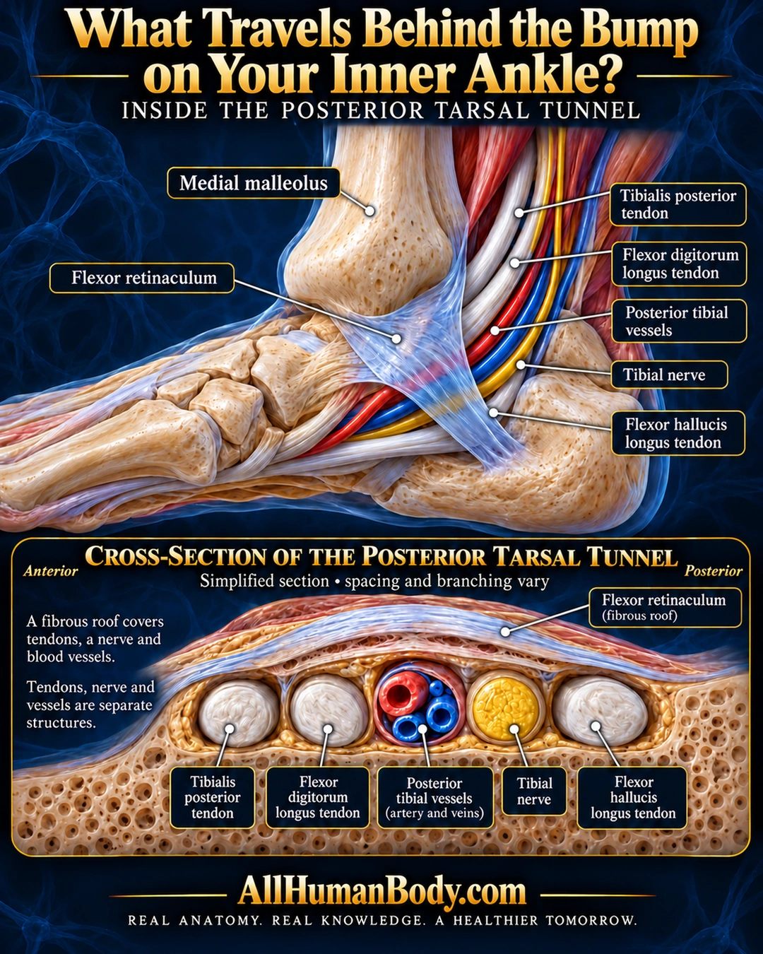
Behind the inner ankle’s medial malleolus, the flexor retinaculum covers the posterior tarsal tunnel. Several tendons, the tibial nerve and posterior tibial vessels pass through as separate structures.
READ THE IMAGE
What the illustration shows
English anatomical poster on midnight navy. A medial ankle cutaway shows the inner ankle bone, with translucent flexor retinaculum bridging over three ivory tendons, red and blue posterior tibial vessels and a gold tibial nerve behind it. Direct labels identify each structure. Below, an anterior-to-posterior simplified section separates tibialis posterior, flexor digitorum longus, vessels, tibial nerve and flexor hallucis longus beneath the fibrous roof. A qualifier states that spacing and branching vary. Gold AllHumanBody.com branding and the full slogan sit below.
How to interpret this poster
- Schematic spacing and branching vary; illustrated tendon and vessel walls are not verified histology. Posterior tunnel only, with no compression test or treatment guidance.
- AI-assisted educational illustration; not physician certification, microscopy, a diagnosis or individualized medical advice.
- Fine annotations need enlargement; the full description provides an accessible text alternative.
- Display the full uncropped illustration; do not trim titles, legends or boundary annotations.
AI-assisted educational illustration, adapted into web-sized images without changing its composition. This is general education, not an individual diagnosis or treatment recommendation.
EVIDENCE / CONTEXT
Sources and what they support
- Primary human cadaveric study, Fine dissection of the tarsal tunnel in 60 cases, Scientific Reports (2017) ↗
Tunnel boundaries and tendon/neurovascular contents; variable nerve branching cautions.