Musculoskeletal / VISUAL EXPLAINER
Your Ear and Knee Use Different Kinds of Cartilage
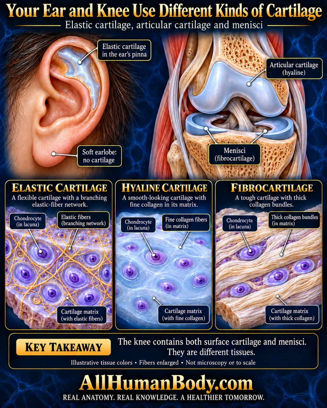
Cartilage types differ in their supporting matrix. The ear’s pinna illustrates elastic cartilage, while the knee has hyaline surface cartilage and fibrocartilage menisci.
READ THE IMAGE
What the illustration shows
English poster titled Your Ear and Knee Use Different Kinds of Cartilage on navy. Left ear has an upper-pinna cartilage window and an unhighlighted soft earlobe labeled no cartilage. Right knee cutaway distinguishes pale blue articular surface lining from two crescent-shaped menisci. Below, three illustrative tissue blocks show elastic cartilage with branching yellow fibers, hyaline cartilage with fine pale collagen, and fibrocartilage with thick ivory bundles. Violet cells in lacunae are identified. A takeaway states the knee has both surface cartilage and menisci. Colors and fibers are illustrative and enlarged, not microscopy or scale. Gold AllHumanBody.com and full slogan below.
How to interpret this poster
- Colors and enlarged fibers are not microscopy or a diagnostic stain guide. Hyaline cartilage is not collagen-free, and the earlobe is not cartilaginous.
- AI-assisted educational illustration; not physician certification, microscopy, a diagnosis or individualized medical advice.
- Fine annotations need enlargement; the full description provides an accessible text alternative.
- Display the full uncropped illustration; do not trim titles, legends or boundary annotations.
AI-assisted educational illustration, adapted into web-sized images without changing its composition. This is general education, not an individual diagnosis or treatment recommendation.
EVIDENCE / CONTEXT
Sources and what they support
- Columbia University histology laboratory ↗
Three classes and matrix-based distinctions; chondrocytes occupy lacunae.
- University of Leeds Histology Guide ↗
Fine collagen versus elastic fiber network versus thick collagen bundles.
- AAOS patient anatomy ↗
Two wedge-shaped fibrocartilage menisci between femur/tibia.
- AAOS patient anatomy ↗
Normal articular cartilage is hyaline.