Special Senses / VISUAL EXPLAINER
How Do Air Spaces Behind Your Ear Connect to the Middle Ear?
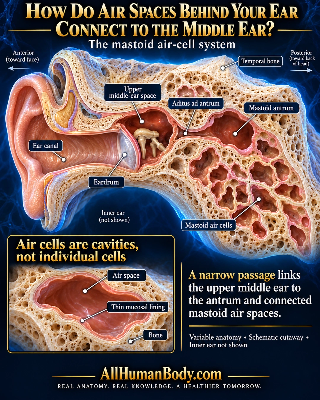
Air-filled cavities in the temporal bone behind the ear connect with the middle-ear space through a narrow passage and the mastoid antrum. Their walls have a mucosal lining; they are not the fluid-filled inner ear.
READ THE IMAGE
What the illustration shows
Navy and gold anatomy poster showing an oblique ivory temporal-bone cutaway. The ear canal and eardrum are on the left. The upper middle-ear cavity communicates through a narrow aditus with a larger mastoid antrum to the right, beside irregular mastoid air spaces. Anterior and posterior orientation labels are shown. A lower inset labels the air space, thin peach mucosal lining and bone. Text says air cells are cavities, not individual cells; inner ear is not shown. Gold AllHumanBody.com branding and the full slogan appear below.
How to interpret this poster
- Cavity branching and lining thickness are schematic and variable, not patient imaging or an exact acoustic network. No mastoiditis diagnosis or pressure-pump claim.
- AI-assisted educational illustration; not physician certification, microscopy, a diagnosis or individualized medical advice.
- Fine annotations need enlargement; the full description provides an accessible text alternative.
- Display the full uncropped illustration; do not trim titles, legends or boundary annotations.
AI-assisted educational illustration, adapted into web-sized images without changing its composition. This is general education, not an individual diagnosis or treatment recommendation.
EVIDENCE / CONTEXT
Sources and what they support
- Acoustical transmission-line model of the middle-ear cavities and mastoid air cells; research paper ↗
Interconnected normal air-filled tympanic cavity, aditus, antrum and mastoid cells within temporal bone.
- Primary human temporal-bone histology study, 15 archived healthy temporal bones ↗
Mucosa examined in tympanic and mastoid/antral regions; supports lining and regional variation, not a specific clinical diagnosis.
Evidence correspondence is limited to the abstract.