Special Senses / VISUAL EXPLAINER
Can Cells in Your Inner Ear Change Their Length?
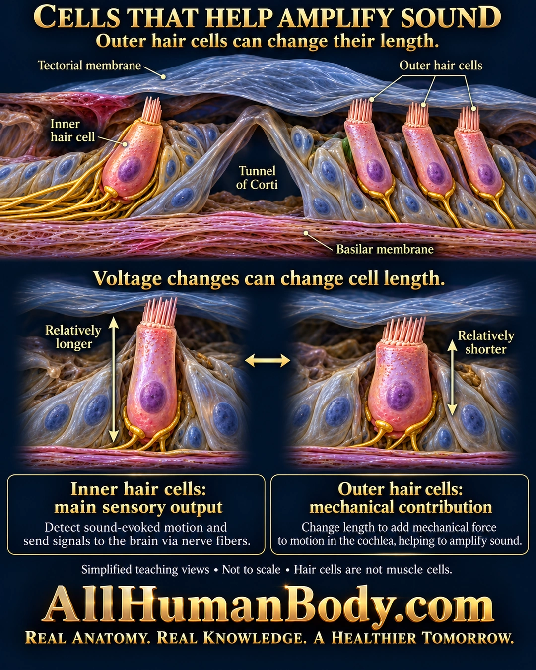
Outer hair cells can change length with membrane-voltage changes, adding a mechanical contribution to cochlear vibration. Inner hair cells provide the main sensory output to separate auditory nerve fibers; hair cells are not muscle cells.
READ THE IMAGE
What the illustration shows
Gold-on-navy poster titled Cells That Help Amplify Sound. A simplified organ of Corti section places one inner hair cell beside the tunnel of Corti and three representative outer hair cells, above the basilar membrane and below the tectorial membrane. The inner bundle has a visible gap below the membrane. Paired enlargements compare relatively longer and shorter states of an outer hair cell with intact membranes and single nuclei. Callouts contrast the main sensory output of inner hair cells with outer cells’ mechanical contribution. The diagram states that hair cells are not muscle cells and is not to scale. Gold AllHumanBody.com branding and full slogan appear below.
How to interpret this poster
- Longer/shorter states, dimensions, bundle shapes and nerve contacts are schematic, not motion recordings.
- No measured displacement, voltage, rate, amplification factor or molecular-motor structure.
- No hearing treatment, regeneration claim or hair-cell axon; visible nerve endings are separate cells.
- AI-assisted educational illustration; not physician certification, microscopy, a diagnosis or individualized medical advice.
- Fine annotations need enlargement; the full description provides an accessible text alternative.
- Display the full uncropped illustration; do not trim titles, legends or boundary annotations.
AI-assisted educational illustration, adapted into web-sized images without changing its composition. This is general education, not an individual diagnosis or treatment recommendation.
EVIDENCE / CONTEXT
Sources and what they support
- MIT OpenCourseWare: Sensory Systems, Fall 2013 — lecture transcript ↗
Outer-cell electromotility/amplification and inner-cell main auditory output.