Special Senses / VISUAL EXPLAINER
Where Does Earwax Come From?
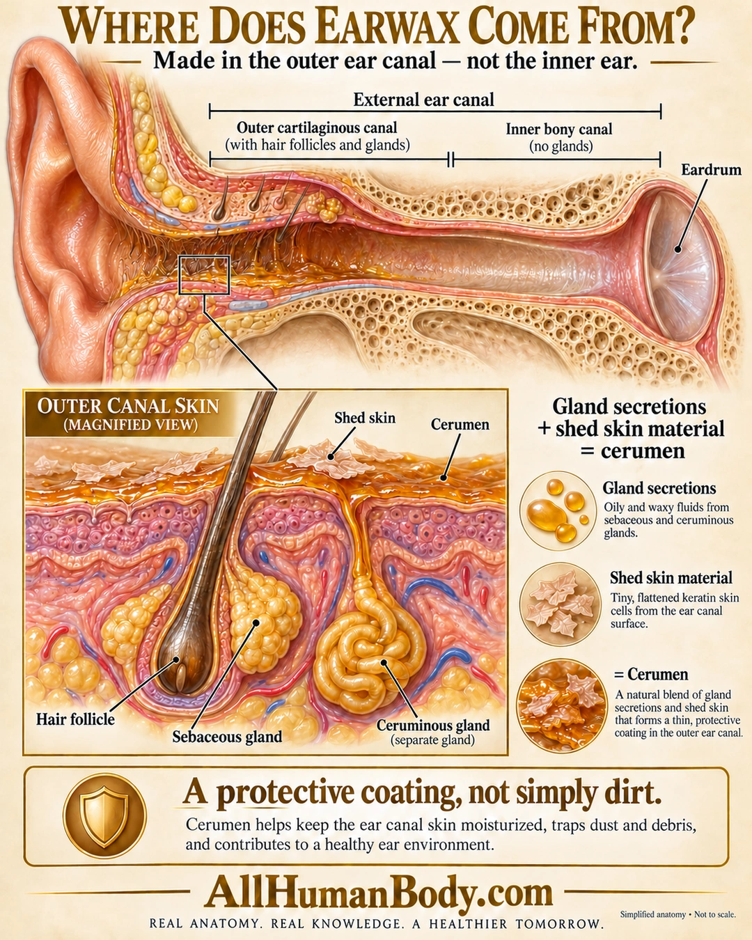
Cerumen forms from gland secretions and shed skin material in the outer ear canal. It helps lubricate canal skin and trap particles; it is not simply dirt or a secretion from the cochlea.
READ THE IMAGE
What the illustration shows
Cream-and-gold educational poster shows an external ear canal from its opening to an intact eardrum. The lateral cartilaginous region contains hairs and glands and a modest amber coating; the medial bony region is clear. A magnified skin inset labels a hair follicle, yellow sebaceous lobules, a coiled ceruminous gland, shed skin flakes and cerumen. A composition panel combines gland secretions with shed skin. The takeaway describes a protective coating, not simply dirt. Structures, coating thickness and duct outlets are schematic, not to scale.
How to interpret this poster
- Canal straightened and widened for teaching, not measured lateral/medial proportions.
- Coating thickness, color and duct outlets are schematic, not a universal wax appearance.
- No removal instructions, irrigation, medication, self-diagnosis or claim all wax is harmless.
- AI-assisted educational illustration; not physician certification, microscopy, a diagnosis or individualized medical advice.
- Fine annotations need enlargement; the full description provides an accessible text alternative.
- Display the full uncropped illustration; do not trim titles, legends or boundary annotations.
AI-assisted educational illustration, adapted into web-sized images without changing its composition. This is general education, not an individual diagnosis or treatment recommendation.
EVIDENCE / CONTEXT
Sources and what they support
- University of Nebraska Medical Center: Unit 1 — Normal Anatomy ↗
Hair and sebaceous/ceruminous glands in lateral cartilaginous canal.
- Mayo Clinic News Network: The scoop on earwax ↗
Normal composition, lubrication and particle trapping.