Special Senses / VISUAL EXPLAINER
How Does Your Inner Ear Sense Head Tilt?
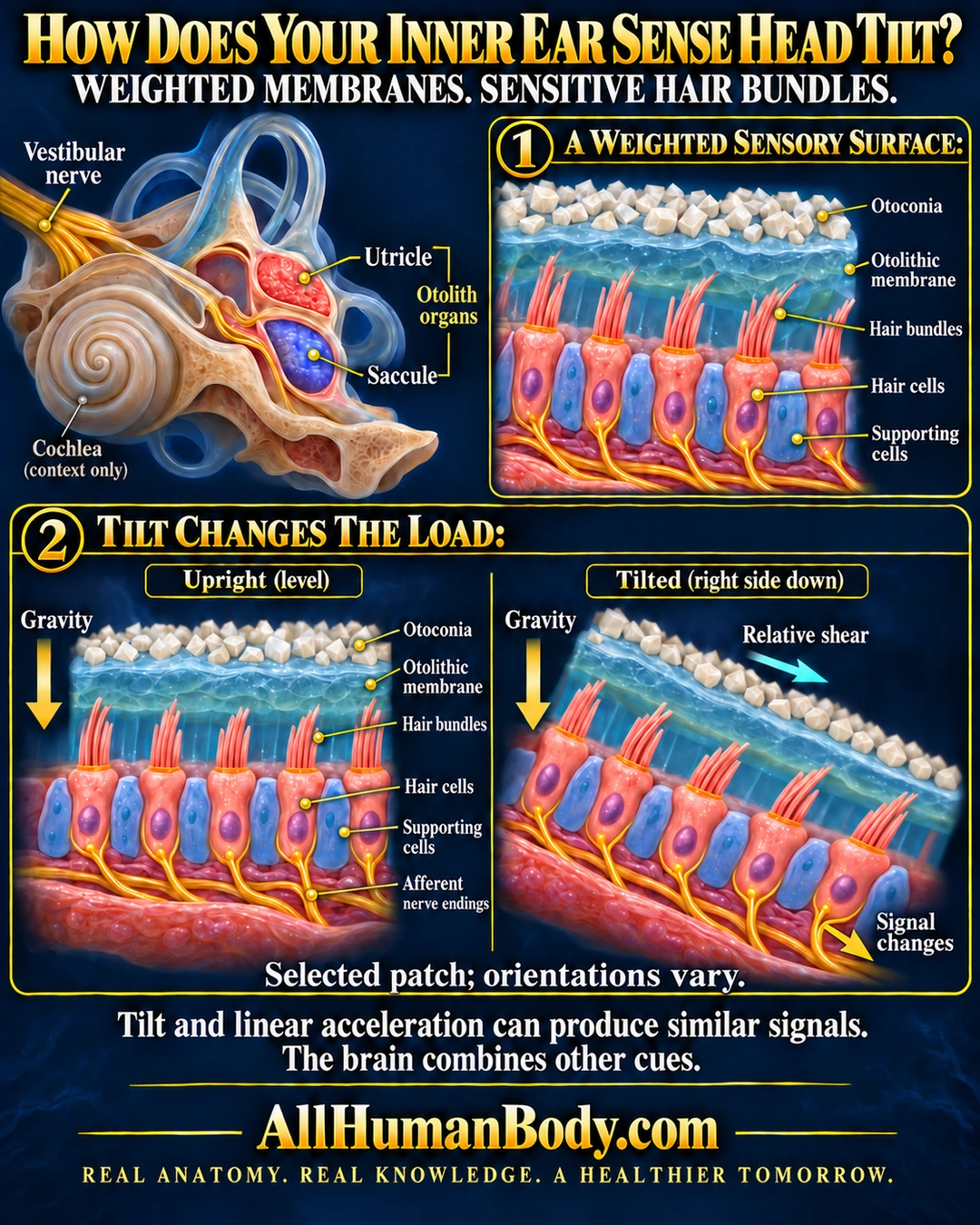
Crystal-loaded membranes in the otolith organs shift relative to sensory hair bundles as gravitational and inertial loads change. Tilt and linear acceleration can produce similar signals, so the brain combines other cues.
READ THE IMAGE
What the illustration shows
Navy-and-gold medical illustration titled “How Does Your Inner Ear Sense Head Tilt?” An inner-ear cutaway labels the vestibular nerve, utricle and saccule, with the cochlea shown for context. A magnified sensory surface shows pale otoconia above a blue otolithic membrane, pink hair bundles extending into the gel from hair cells, blue supporting cells and yellow nerve endings beneath. Below, matched upright and right-side-down sections compare a selected local patch. Both gravity arrows point straight down; a separate cyan arrow shows relative shear down the tilted membrane, and a yellow arrow points outward along the nerve route beside “Signal changes.” The figure states “Selected patch; orientations vary” and explains that tilt and linear acceleration can produce similar signals, so the brain combines other cues. Footer: AllHumanBody.com and “REAL ANATOMY. REAL KNOWLEDGE. A HEALTHIER TOMORROW.”
How to interpret this poster
- Selected local patch, not a whole-organ polarity atlas or exact head-angle decoder.
- The final image does not identify a kinocilium; no named-polarity increase is claimed.
- The linked anchor supplies vestibular structures and interfaces rather than the full tilt comparison.
- AI-assisted educational illustration; not physician certification, microscopy, a diagnosis or individualized medical advice.
- Fine annotations need enlargement; the full description provides an accessible text alternative.
AI-assisted educational illustration, adapted into web-sized images without changing its composition. This is general education, not an individual diagnosis or treatment recommendation.
EVIDENCE / CONTEXT
Sources and what they support
- Balance Disorders ↗
Normal otolith organs, calcium-carbonate grains, sensory bundles and integrated balance.
- Vestibular System: Structure and Function ↗
Macular architecture, directional response and distinction from semicircular-duct receptors.
- Vestibular perception and action employ qualitatively different mechanisms. I. Frequency response of VOR and perceptual responses during Translation and Tilt ↗
Human experiments showing equivalent otolith cues in different motions and contribution of canal cues to perception.
Evidence correspondence is limited to the abstract.