Special Senses / VISUAL EXPLAINER
Is Your Eardrum Just One Layer of Skin?
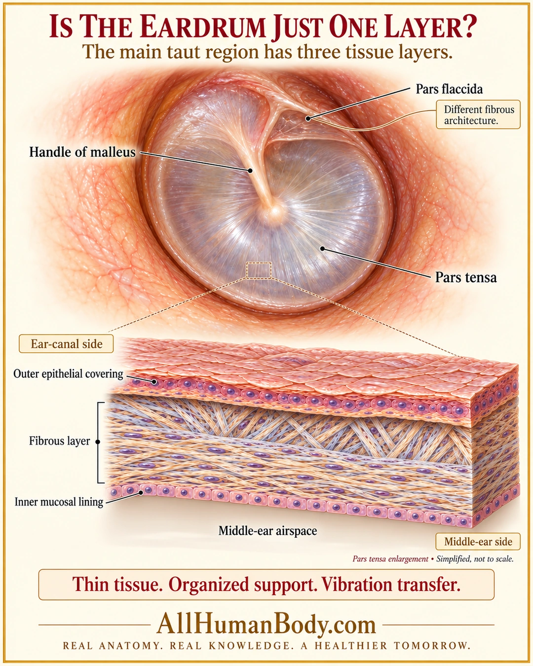
The main taut part of the eardrum combines an outer epithelial covering, fibrous connective support and an inner mucosal lining. This layered boundary transmits vibration between the ear canal and middle-ear mechanical system; the small pars flaccida differs.
READ THE IMAGE
What the illustration shows
Cream-and-gold atlas poster shows a translucent eardrum with its main pars tensa, small superior pars flaccida and attached malleus handle. A magnified pars-tensa slab depicts outer epithelium facing the ear canal, a central collagen layer and a thin inner epithelial lining facing empty middle-ear airspace. A note distinguishes pars flaccida architecture. The takeaway links organized tissue support with vibration transfer. Layer thickness, fiber arrangements and cellular shape are illustrative, not measured or diagnostic.
How to interpret this poster
- Layer thickness, fiber arrangement and cell shapes are illustrative, not a diagnostic otoscopic or histologic image.
- No exact radial/circular reconstruction, cell counts or side-specific orientation.
- No repair advice, normal-thickness number or direct eardrum-to-inner-ear contact.
- AI-assisted educational illustration; not physician certification, microscopy, a diagnosis or individualized medical advice.
- Fine annotations need enlargement; the full description provides an accessible text alternative.
- Display the full uncropped illustration; do not trim titles, legends or boundary annotations.
AI-assisted educational illustration, adapted into web-sized images without changing its composition. This is general education, not an individual diagnosis or treatment recommendation.
EVIDENCE / CONTEXT
Sources and what they support
- University of Nebraska Medical Center: Unit 1 — Normal Anatomy ↗
Tympanic outer epithelium, fibrous support and inner mucosa.
- MedlinePlus Medical Encyclopedia: Tympanic membrane ↗
Connective membrane with outer skin and inner mucosa.