Digestive / VISUAL EXPLAINER
How Does a Liver Cell Keep Blood Exchange Separate from Bile?
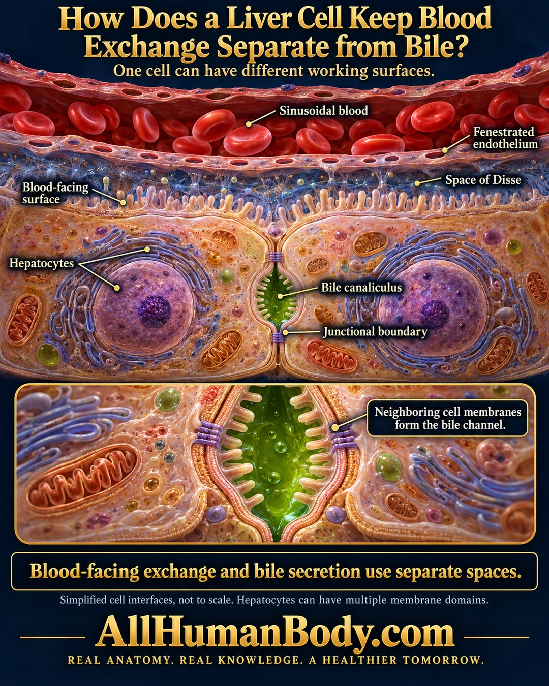
A hepatocyte exchanges material at blood-facing surfaces and secretes bile into distinct canalicular spaces formed with neighboring cells. Junctions help maintain this separation; one cell can contribute to more than one bile-facing domain.
READ THE IMAGE
What the illustration shows
English medical poster showing two adjacent hepatocytes beneath a blood sinusoid. Red blood cells remain inside the upper red lumen. A thin fenestrated endothelial lining separates it from the blue-tinted space of Disse and the hepatocytes’ microvilli. A small green bile canaliculus lies between the two cells, with junctional contacts beside its lumen. An enlarged lower inset shows neighboring cell membranes and microvilli around the green bile channel. The takeaway says blood-facing exchange and bile secretion use separate spaces. The image notes that interfaces are simplified and hepatocytes can have multiple membrane domains. Gold AllHumanBody.com branding and the complete project slogan appear below.
How to interpret this poster
- Selected interfaces only, not a complete three-dimensional canalicular network or exact pore dimensions.
- Sinusoidal endothelium and the space of Disse intervene between blood and hepatocytes; red cells remain in the blood lumen.
- Rat-cell culture supports junctional organization, not quantitative human measurements; no detox or treatment claim.
- AI-assisted educational illustration; not physician certification, microscopy, a diagnosis or individualized medical advice.
- Fine annotations need enlargement; the full description provides an accessible text alternative.
- Display the full uncropped illustration; do not trim titles, legends or boundary annotations.
AI-assisted educational illustration, adapted into web-sized images without changing its composition. This is general education, not an individual diagnosis or treatment recommendation.
EVIDENCE / CONTEXT
Sources and what they support
- University of Leeds Histology Guide: Hepatocytes ↗
Sinusoidal lining, exchange space, microvilli and intercellular canaliculi.
- Structural and functional hepatocyte polarity and liver disease ↗
Normal basal and potentially multiple apical domains.
Evidence correspondence is limited to the abstract. - Primary cultures of rat hepatocytes as a model system of canalicular development, biliary secretion, and intrahepatic cholestasis. I. Distribution of filipin-cholesterol complexes during de novo formation of bile canaliculi ↗
Rat-culture canaliculi sealed by tight junctions.
Evidence correspondence is limited to the abstract.