Digestive / VISUAL EXPLAINER
How Does Blood Exchange Material With Liver Cells?
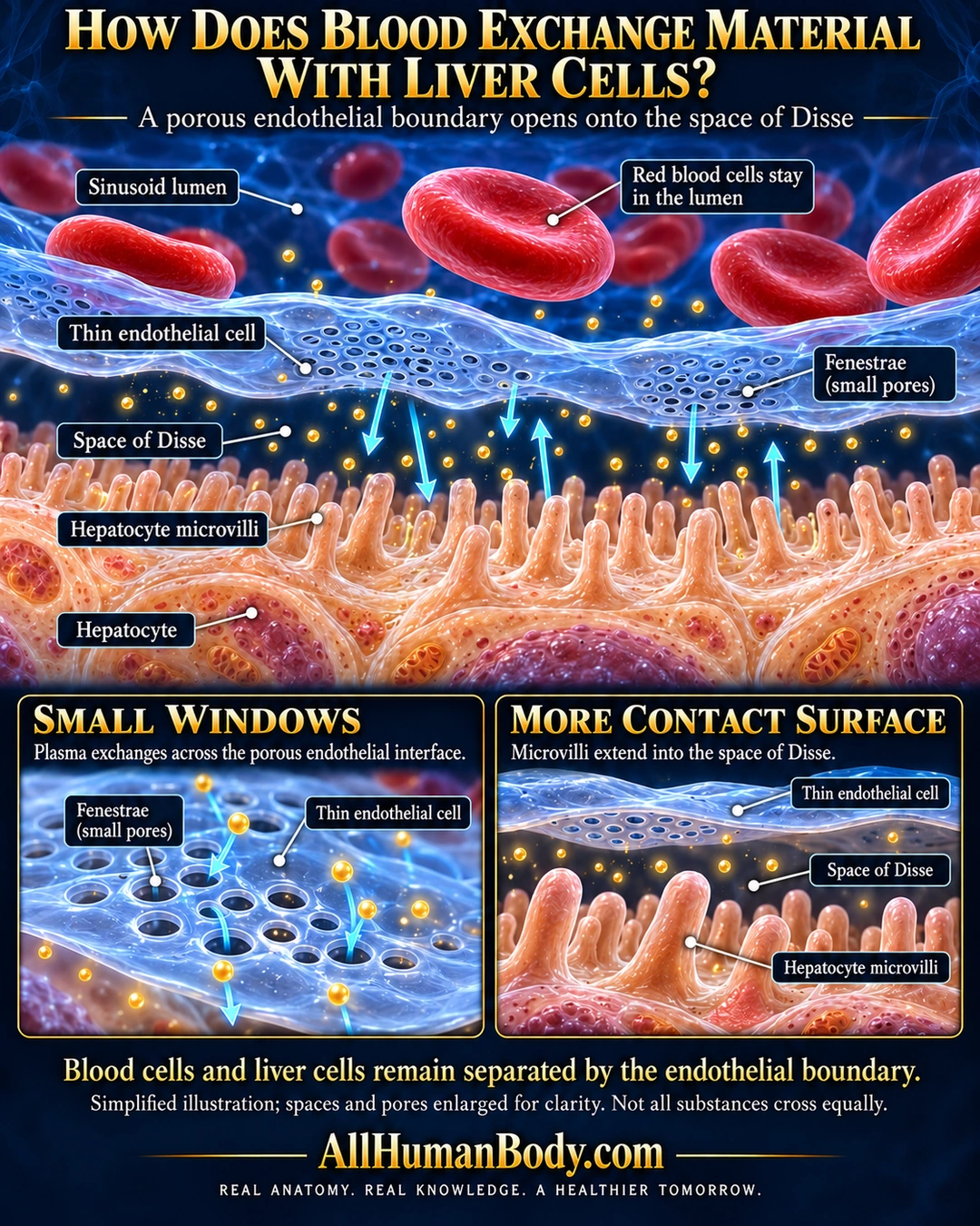
Liver sinusoidal endothelium forms a thin porous exchange boundary. Plasma constituents can reach the space of Disse and the microvilli of hepatocytes, while blood cells remain within the sinusoidal lumen; substances do not all cross equally.
READ THE IMAGE
What the illustration shows
Navy and gold liver exchange poster. Red blood cells occupy the upper blue sinusoid lumen above a thin blue endothelial sheet containing clusters of small pores. Gold dots and cyan arrows illustrate exchange through the pores into the space of Disse. Peach hepatocyte microvilli project upward into that space from liver cells below. Two lower insets enlarge the endothelial pores and the gap between the endothelium and microvilli. Labels identify each boundary. A qualifier says spaces and pores are enlarged and not all substances cross equally. Gold AllHumanBody.com branding and full slogan sit on a plain navy footer.
How to interpret this poster
- Pores, gap thickness, microvilli and arrows are enlarged and qualitative, not measured diameters, clearance rates or flow.
- Other sinusoidal cells, matrix and membrane transport steps are omitted for focus, not absent in tissue.
- This is not a kidney filter, unrestricted sieve or detox regimen.
- AI-assisted educational illustration; not physician certification, microscopy, a diagnosis or individualized medical advice.
- Fine annotations need enlargement; the full description provides an accessible text alternative.
- Display the full uncropped illustration; do not trim titles, legends or boundary annotations.
AI-assisted educational illustration, adapted into web-sized images without changing its composition. This is general education, not an individual diagnosis or treatment recommendation.
EVIDENCE / CONTEXT
Sources and what they support
- University of Leeds Histology: Liver hepatocytes ↗
Sinusoidal endothelial pores, plasma access to Disse, blood-cell exclusion and hepatocyte microvilli.