Digestive / VISUAL EXPLAINER
Are All Three Tubes in a Liver Portal Tract Blood Vessels?
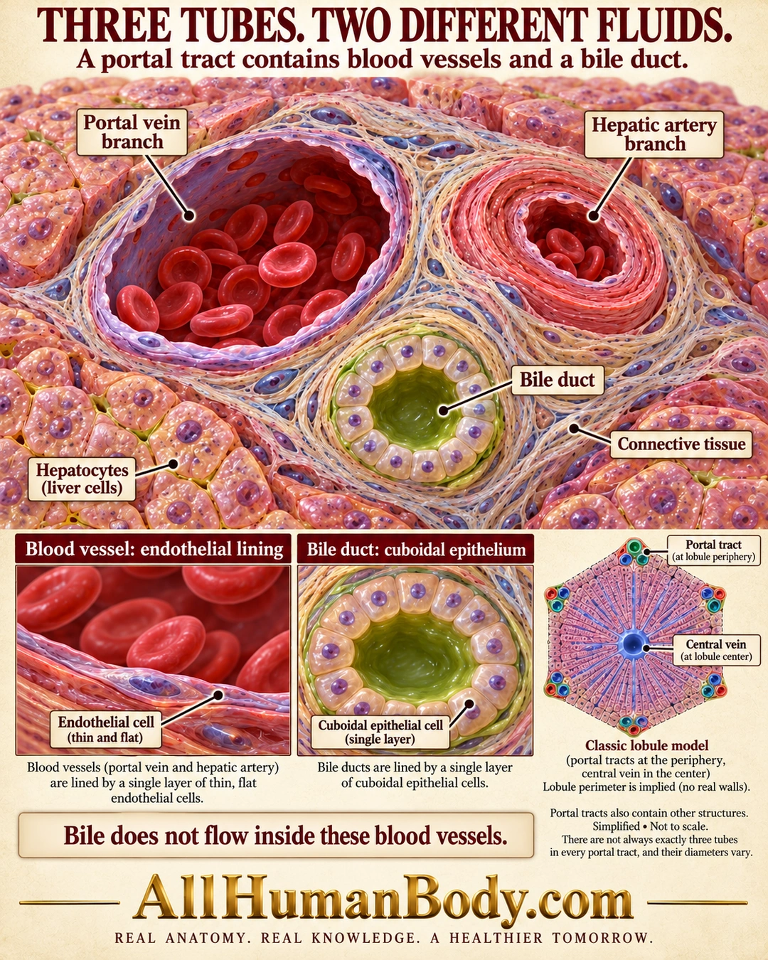
A liver portal tract brings portal venous and hepatic arterial branches close to a bile duct, but their contents remain separate. Small bile ducts have cuboidal epithelium rather than the thin endothelial lining of blood vessels; the classical lobule places portal tracts peripherally and a central vein centrally.
READ THE IMAGE
What the illustration shows
Ivory medical poster titled Three Tubes. Two Different Fluids. A portal-tract tissue cutaway shows a large thin-walled portal vein branch and a smaller thicker-walled hepatic artery branch, both containing red cells, beside a separate greenish bile duct with a single ring of cuboidal epithelial cells and no red cells. Connective tissue surrounds the tubes; hepatocytes lie outside. Lower enlargements contrast thin flat endothelium with cuboidal duct epithelium. A classic lobule model places a central vein centrally and portal tracts around the periphery. Text notes other portal-tract structures and variable diameters. Gold AllHumanBody.com branding and the complete slogan form the footer.
How to interpret this poster
- Portal triad is shorthand: tracts contain additional structures and sections do not always show exactly three tubes.
- Relative calibers, cell counts and color are illustrative; no literal thick hexagonal walls around every human lobule.
- No diagnostic staining, bile-to-blood continuity or detoxification claim.
- AI-assisted educational illustration; not physician certification, microscopy, a diagnosis or individualized medical advice.
- Fine annotations need enlargement; the full description provides an accessible text alternative.
- Display the full uncropped illustration; do not trim titles, legends or boundary annotations.
AI-assisted educational illustration, adapted into web-sized images without changing its composition. This is general education, not an individual diagnosis or treatment recommendation.
EVIDENCE / CONTEXT
Sources and what they support
- AASLD Pathology Pearls: Normal Liver Histology 101 ↗
Portal-tract tubes, luminal linings and classical lobule organization.