Cardiovascular / VISUAL EXPLAINER
Which Layers Surround Your Heart?
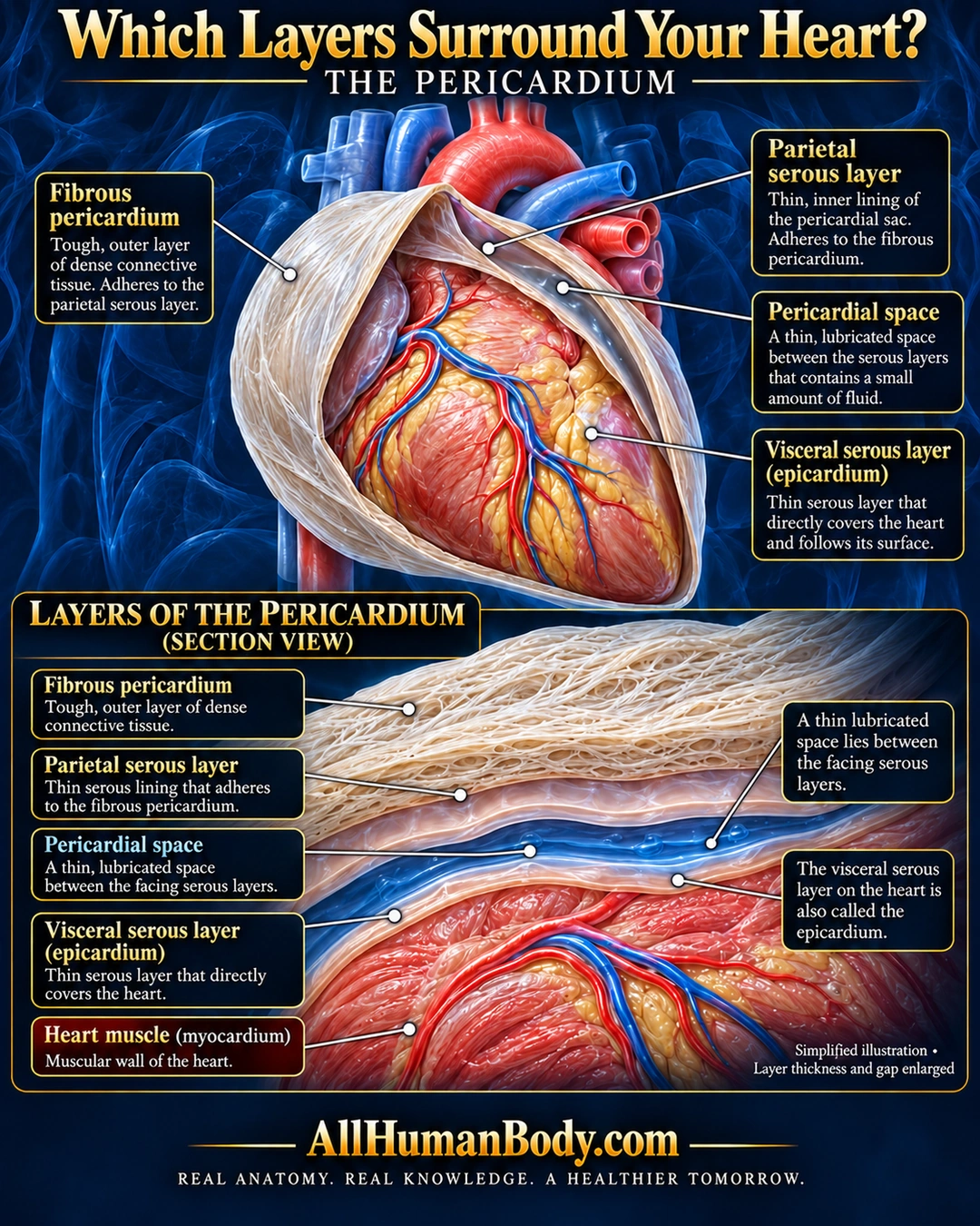
The pericardium combines an outer fibrous layer with parietal and visceral serous layers. The visceral layer on the heart is also called epicardium; a thin lubricated space lies between the facing serous surfaces.
READ THE IMAGE
What the illustration shows
English medical poster on navy background. A partly opened pale fibrous sac surrounds an external heart. Labels identify fibrous pericardium, its adherent parietal serous lining, pericardial space and visceral serous layer or epicardium on the heart. A lower enlarged section orders ivory fibrous tissue, pale parietal membrane, blue fluid space, pale visceral membrane and red heart muscle. Gap and layer thickness are enlarged for teaching. Blue is illustrative, not actual fluid color. Gold AllHumanBody.com and full slogan below.
How to interpret this poster
- Gap and layer thickness are enlarged, with blue as symbolic fluid color. Not a giant fluid bag, blood-filled cavity or disease/drainage guide; fine vessels are contextual.
- AI-assisted educational illustration; not physician certification, microscopy, a diagnosis or individualized medical advice.
- Fine annotations need enlargement; the full description provides an accessible text alternative.
- Display the full uncropped illustration; do not trim titles, legends or boundary annotations.
AI-assisted educational illustration, adapted into web-sized images without changing its composition. This is general education, not an individual diagnosis or treatment recommendation.
EVIDENCE / CONTEXT
Sources and what they support
- OpenStax anatomy textbook ↗
All three layer and fluid-space claims.