Respiratory / VISUAL EXPLAINER
How Can Pneumonia Fill the Lungs’ Air Sacs?
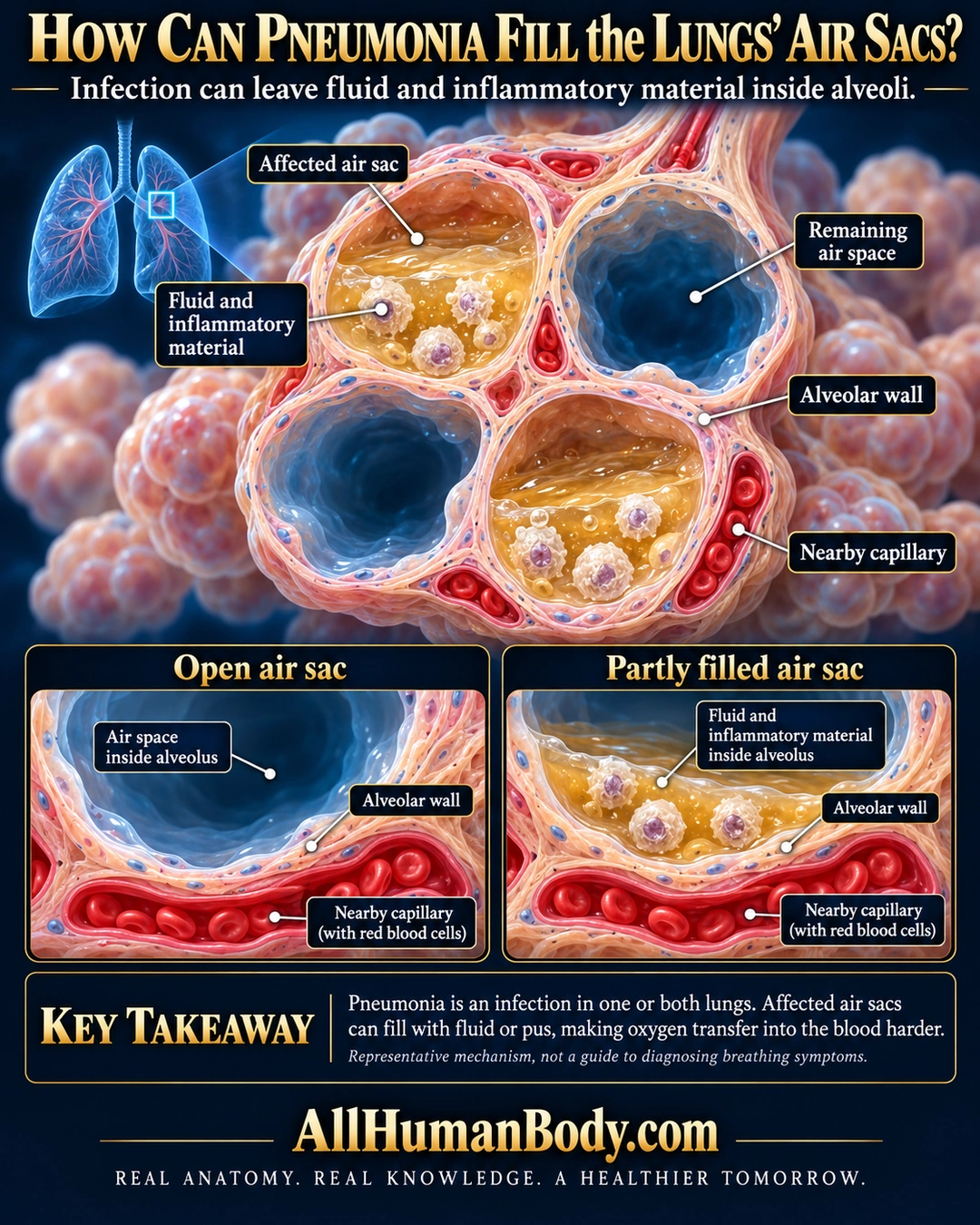
Pneumonia is an infection in one or both lungs. Affected air sacs can fill with fluid and inflammatory material, making oxygen transfer into blood harder.
READ THE IMAGE
What the illustration shows
English educational poster titled “How Can Pneumonia Fill the Lungs’ Air Sacs?” on midnight navy. A small blue lung locator connects to a large cutaway of four neighboring air sacs. Two lumens contain golden fluid and pale inflammatory cells; two remain dark blue and open. Peach alveolar walls surround each cavity. Separate red capillaries in the walls contain red blood cells. Leaders identify an affected air sac, fluid and inflammatory material, remaining air space, alveolar wall and nearby capillary. Lower open-versus-partly-filled windows show the epithelial boundary between air-space contents and capillary blood. Text says pneumonia is an infection in one or both lungs, and affected air sacs can fill with fluid or pus, making oxygen transfer harder. A qualifier says this is a representative mechanism, not a guide to diagnosing breathing symptoms. Gold AllHumanBody.com and the full slogan sit on a plain navy footer.
How to interpret this poster
- Representative pattern, not organism identification, severity, universal distribution or all exchange stopping. No breathing-symptom assessment or treatment.
- AI-assisted educational illustration; not physician certification, a diagnosis or individualized medical advice.
- Fine annotations require enlargement; preserve the full uncropped illustration and its qualifiers.
AI-assisted educational illustration, adapted into web-sized images without changing its composition. This is general education, not an individual diagnosis or treatment recommendation.
EVIDENCE / CONTEXT
Sources and what they support
- NHLBI: Pneumonia ↗
Infection and possible alveolar fluid or pus.
- American Lung Association: Pneumonia ↗
Alveolar filling and impaired oxygen transfer.