Respiratory / VISUAL EXPLAINER
What Passes Through the Inner Doorway of a Lung?
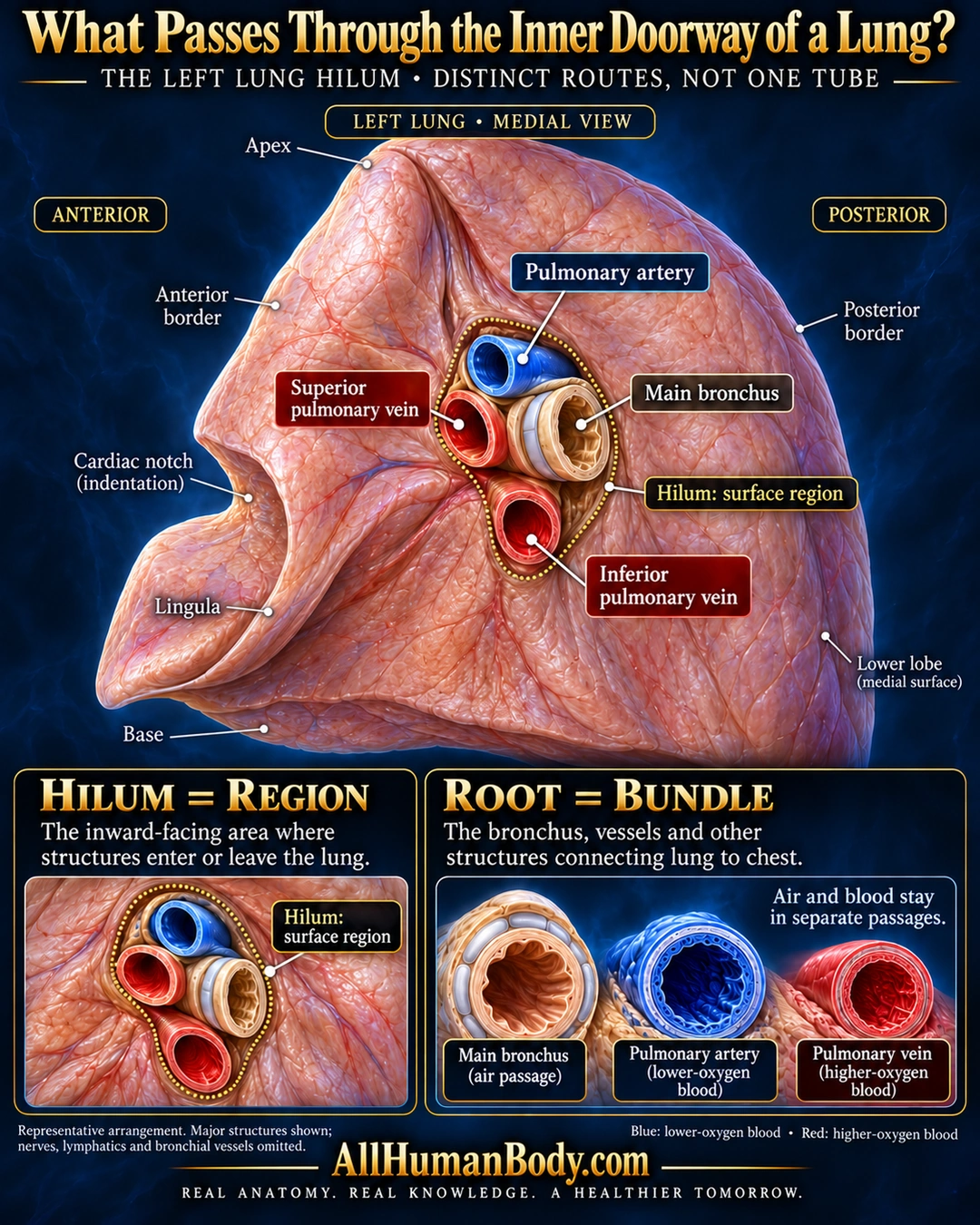
The hilum is the inward-facing surface region where the lung’s root passes. The root includes a bronchus and distinct blood vessels, so air and blood use separate passages rather than one shared tube.
READ THE IMAGE
What the illustration shows
English medical poster titled “What Passes Through the Inner Doorway of a Lung?” A light-peach left lung is shown from its medial side on a dark navy and cyan background, with anterior at the left and posterior at the right. Labels identify apex, anterior and posterior borders, cardiac notch, lingula, base and lower-lobe medial surface. A dotted gold outline marks the hilum. Four distinct cut openings show a blue pulmonary artery above an ivory cartilage-ringed main bronchus, a red superior pulmonary vein anterior to it and a red inferior pulmonary vein below. The lower-left panel repeats the outlined surface region, while the lower-right panel shows separate bronchial and vascular cross-sections and explains that air and blood remain in separate passages. Text distinguishes the hilum region from the root bundle and notes that this representative arrangement shows major structures only, omitting nerves, lymphatics and bronchial vessels. Blue and red indicate relatively lower- and higher-oxygen blood. Gold AllHumanBody.com branding and the full slogan appear at the bottom.
How to interpret this poster
- Representative left-sided major structures only, not a universal right/left arrangement or complete root inventory. Colors indicate relative oxygenation; generated wall textures are not histology.
- AI-assisted educational illustration; not physician certification, microscopy, a diagnosis or individualized medical advice.
- Fine annotations need enlargement; the full description provides an accessible text alternative.
- Display the full uncropped illustration; do not trim titles, legends or boundary annotations.
AI-assisted educational illustration, adapted into web-sized images without changing its composition. This is general education, not an individual diagnosis or treatment recommendation.
EVIDENCE / CONTEXT
Sources and what they support
- Washington State University Elson S. Floyd College of Medicine anatomy laboratory guide ↗
Medial root location; pulmonary arteries, veins, bronchi and hilar nodes; right/left arrangement differs.
- Texas Tech University Health Sciences Center El Paso gross anatomy teaching resource ↗
Hilum versus root, posterior bronchus, anterior/inferior veins, left pulmonary artery superior to bronchus; pulmonary relative oxygenation color convention.