Respiratory / VISUAL EXPLAINER
How Pulmonary Fibrosis Changes Lung Tissue 🫁
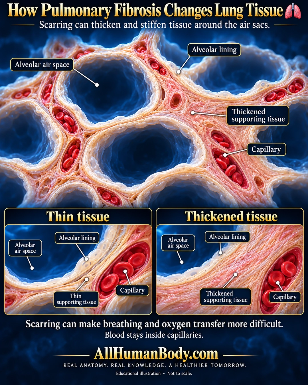
Pulmonary fibrosis is scarring in lung tissue. Tissue around air sacs can thicken and stiffen, making breathing and oxygen transfer more difficult.
READ THE IMAGE
What the illustration shows
Medical illustration on a navy background titled How Pulmonary Fibrosis Changes Lung Tissue, with a lungs symbol. Large empty blue alveolar air spaces are separated by continuous ivory linings and thick peach supporting tissue. Separate capillary channels within the partitions contain red blood cells. Labels identify air space, lining, supporting tissue and capillary. Two lower panels compare a thin tissue barrier with a thickened one, while keeping air and capillary blood separate. Text states that scarring can make breathing and oxygen transfer more difficult. Gold AllHumanBody.com and the full slogan appear below. Educational illustration, not to scale.
How to interpret this poster
- Generated separation exaggerated, not measured diffusion distance or a universal histological pattern. No all-causes-unknown inference, prognosis or treatment.
- AI-assisted educational illustration; not physician certification, a diagnosis or individualized medical advice.
- Fine annotations require enlargement; preserve the full uncropped illustration and its qualifiers.
AI-assisted educational illustration, adapted into web-sized images without changing its composition. This is general education, not an individual diagnosis or treatment recommendation.
EVIDENCE / CONTEXT
Sources and what they support
- NHLBI: Idiopathic pulmonary fibrosis ↗
Thick/stiff scarred tissue around alveoli; IPF only an example.
- NHLBI: Interstitial lung diseases ↗
Supporting tissue change and oxygen-transfer difficulty.