Respiratory / VISUAL EXPLAINER
How Food and Blood Cross Your Breathing Muscle
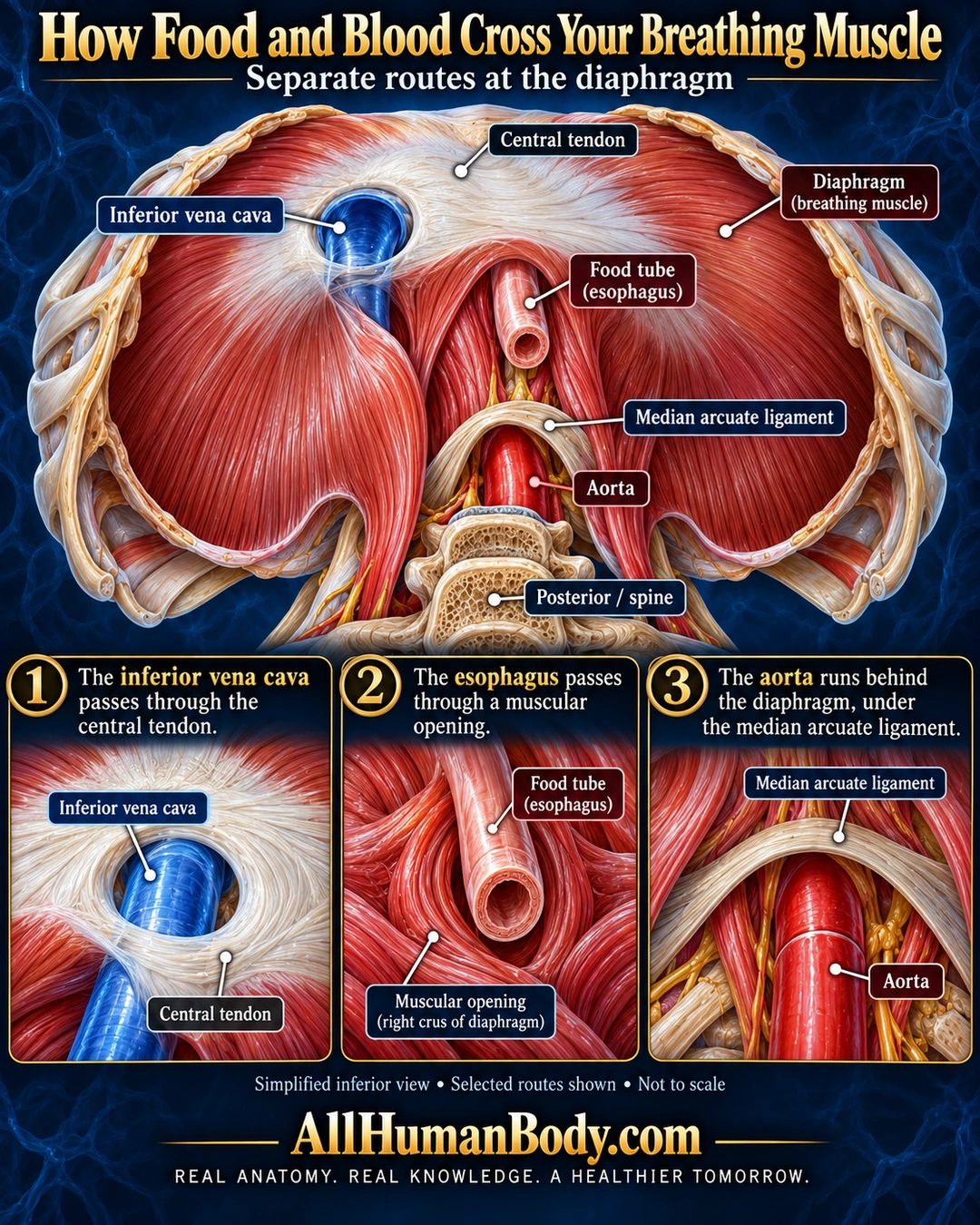
Major vessels and the food tube use different routes at the diaphragm. The vena cava crosses the central tendon, the esophagus passes through muscle, and the aorta runs behind the diaphragm beneath a ligament.
READ THE IMAGE
What the illustration shows
English poster titled How Food and Blood Cross Your Breathing Muscle on navy. An inferior oblique diaphragm view shows pale central tendon, red muscle, blue inferior vena cava through tendon, pink esophagus through a muscular opening, and red aorta posteriorly against the spine beneath a pale median arcuate ligament. Three lower closeups repeat these distinct routes, each with precise pointers and short text. Posterior spine is identified. Qualifier says simplified inferior view, selected routes shown, not to scale. Gold AllHumanBody.com and full slogan below.
How to interpret this poster
- Selected passageways only, not every crossing structure; no rigid vertebral levels or surgical precision. The view is simplified and not to scale.
- AI-assisted educational illustration; not physician certification, microscopy, a diagnosis or individualized medical advice.
- Fine annotations need enlargement; the full description provides an accessible text alternative.
- Display the full uncropped illustration; do not trim titles, legends or boundary annotations.
AI-assisted educational illustration, adapted into web-sized images without changing its composition. This is general education, not an individual diagnosis or treatment recommendation.
EVIDENCE / CONTEXT
Sources and what they support
- Texas Tech anatomy teaching explanation ↗
Caval tendon opening, esophageal route, aorta deep to median arcuate ligament.
- Elsevier Complete Anatomy ↗
Distinct major passageways and relevant muscle/tendon relationships.