Respiratory / VISUAL EXPLAINER
How Do Your Airways Move Mucus Out?
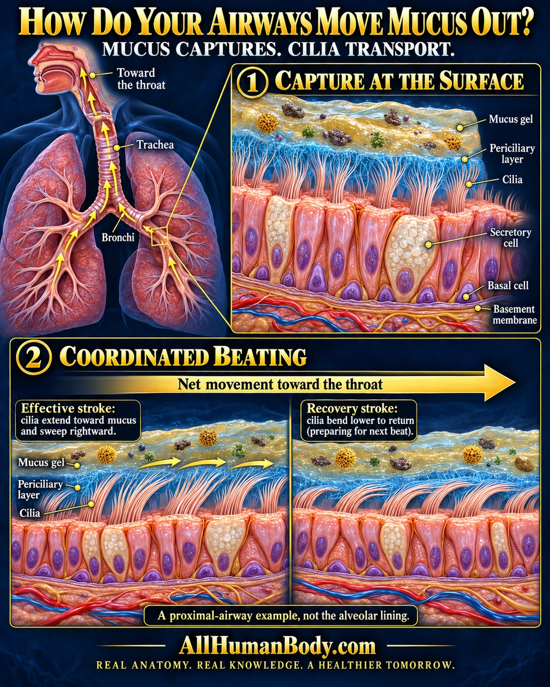
Airway mucus catches some inhaled material. Coordinated ciliary beating helps move it toward the throat, where it can be swallowed or expelled; this lining is different from the gas-exchange surface of alveoli.
READ THE IMAGE
What the illustration shows
Educational poster titled “How Do Your Airways Move Mucus Out?” on a navy background. At left, gold arrows follow the bronchi into the trachea and upward toward the throat. A large cutaway labeled “Capture at the surface” shows particles caught in gold mucus gel above a blue periciliary layer surrounding coral cilia. Beneath are tall ciliated cells, pale granule-filled secretory cells, and small basal cells within one epithelial sheet above the basement membrane. The lower section, “Coordinated beating,” compares higher, rightward-sweeping cilia during the effective stroke with lower-bent cilia during recovery. A broad rightward arrow indicates net movement toward the throat. A note identifies this as a proximal-airway example, not the alveolar lining. Footer: AllHumanBody.com; “Real anatomy. Real knowledge. A healthier tomorrow.”
How to interpret this poster
- A local proximal-airway example, not a uniform mucus blanket or an alveolar lining.
- Beat panels are simplified snapshots, not a measured wave or a claim of zero backward movement.
- Fine labels require enlargement; preserve the full image because original margin aspirations were not all met.
- AI-assisted educational illustration; not physician certification, microscopy, a diagnosis or individualized medical advice.
- Fine annotations need enlargement; the full description provides an accessible text alternative.
AI-assisted educational illustration, adapted into web-sized images without changing its composition. This is general education, not an individual diagnosis or treatment recommendation.
EVIDENCE / CONTEXT
Sources and what they support
- Primary Ciliary Dyskinesia ↗
Normal introduction explaining cilia and mucus removal; disease and treatment content excluded.
- Physiology and pathophysiology of human airway mucus ↗
Distinct mucus and periciliary compartments, effective/recovery strokes and proximal net transport.