Respiratory / VISUAL EXPLAINER
Are Your Left and Right Lungs Mirror Images?
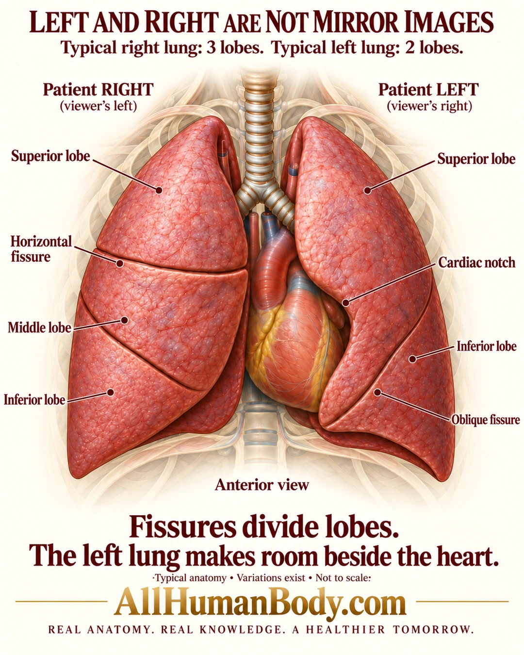
The typical right lung has superior, middle and inferior lobes; the typical left has superior and inferior lobes. Fissures divide these regions, and a cardiac notch contributes to the left lung’s asymmetric contour beside the heart.
READ THE IMAGE
What the illustration shows
An anterior anatomical illustration compares the two lungs. The patient right lung appears on the viewer left and is labeled with superior, middle and inferior lobes and a horizontal fissure. The patient left lung on the viewer right has superior and inferior lobes, an oblique fissure and cardiac notch next to the visible central heart. A trachea and primary bronchi lie above the heart, with faint ribs behind. Text states that the typical right lung has three lobes and left two, fissures divide lobes, and variations exist. Gold AllHumanBody.com branding and the full slogan sit below. Simplified anatomy, not to scale.
How to interpret this poster
- Typical anatomy with normal variation, not a diagnostic or surgical map.
- Posterior shadows are depth cues, not extra labeled lobes; fissure completeness, sizes, surface detail and background ribs are schematic.
- No claim that the heart mechanically removes a lobe or that the lingula is a separate third left lobe.
- AI-assisted educational illustration; not physician certification, microscopy, a diagnosis or individualized medical advice.
- Fine annotations need enlargement; the full description provides an accessible text alternative.
- Display the full uncropped illustration; do not trim titles, legends or boundary annotations.
AI-assisted educational illustration, adapted into web-sized images without changing its composition. This is general education, not an individual diagnosis or treatment recommendation.
EVIDENCE / CONTEXT
Sources and what they support
- Anatomy and Physiology 2e: 22.2 The Lungs ↗
Typical lobe counts, fissures, cardiac notch and asymmetric shape; preserved directly opened gross anatomy paragraphs.
- NHLBI: How the Lungs Work — The Respiratory System ↗
Typical three right and two left lobes and heart-related size difference.