Respiratory / VISUAL EXPLAINER
How Do Your Lungs Follow Your Moving Chest?
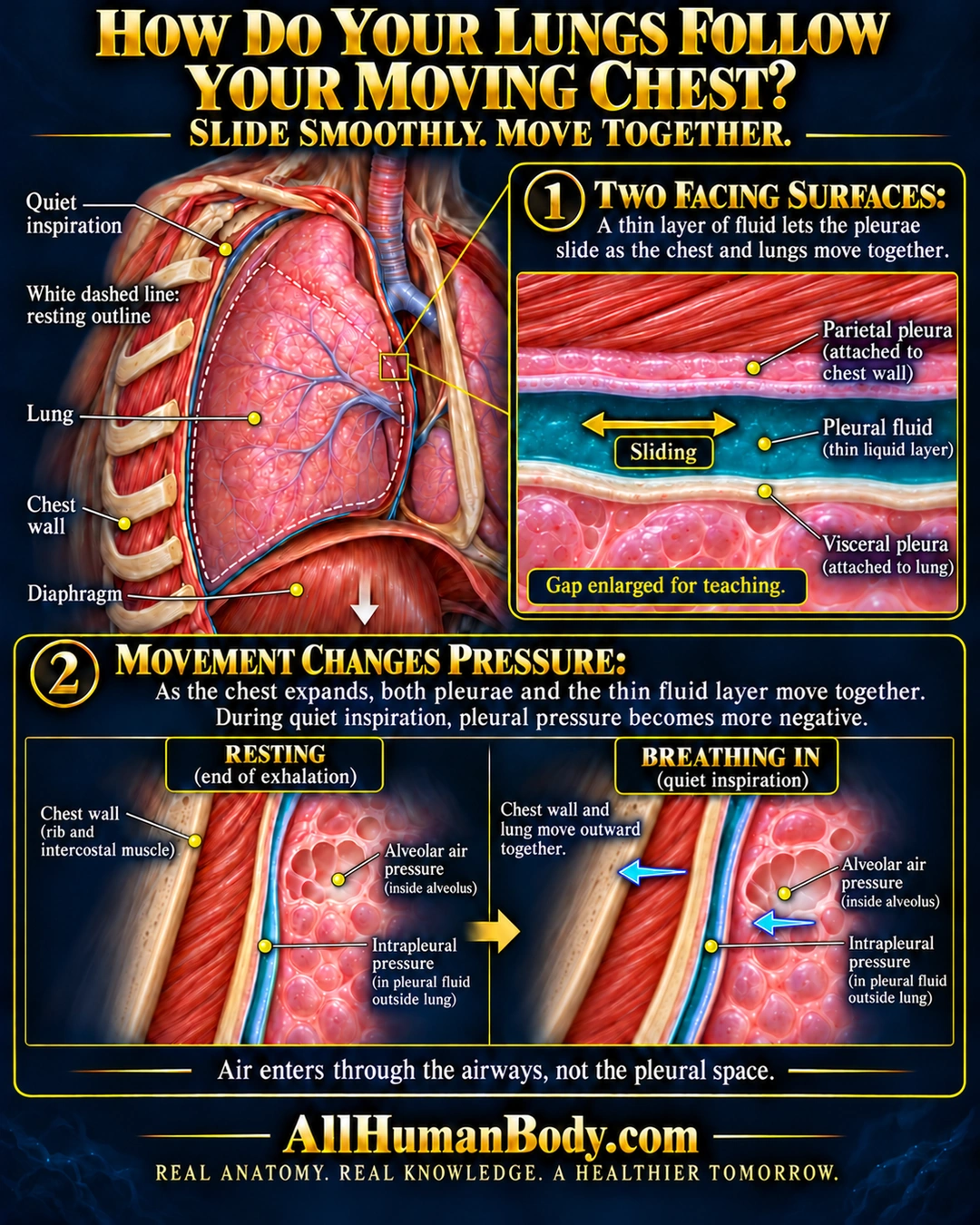
The lung and chest wall move together while their pleural surfaces can slide across a thin fluid interface. During quiet spontaneous inspiration, chest expansion changes pressure relationships; breathing air enters through the airways, not the pleural space.
READ THE IMAGE
What the illustration shows
Navy-and-gold educational anatomy poster titled “How Do Your Lungs Follow Your Moving Chest?” with the subtitle “Slide smoothly. Move together.” A thoracic cutaway shows a pink lung beside the chest wall and above the diaphragm. A smaller white dashed contour marks the resting outline; a downward arrow illustrates diaphragm descent during quiet inspiration. An enlarged horizontal inset shows the parietal pleura attached to the chest wall, teal pleural fluid between the surfaces, and visceral pleura attached to the lung. A double-headed arrow runs along the interface to indicate sliding. A note reads “Gap enlarged for teaching.” Below, matched resting and breathing-in details distinguish alveolar air pressure inside an airspace from intrapleural pressure in the liquid outside the lung. Two leftward arrows in the breathing-in panel show chest wall and lung moving outward together. Text states that pleural pressure becomes more negative during quiet inspiration and that air enters through the airways, not the pleural space. Footer: AllHumanBody.com. Fine labels are best read enlarged.
How to interpret this poster
- The fluid gap is enlarged for teaching, not a normal measured thickness.
- Quiet spontaneous breathing only, not forced maneuvers, ventilation or disease.
- The linked section teaches the pleural interface rather than the full pressure sequence.
- AI-assisted educational illustration; not physician certification, microscopy, a diagnosis or individualized medical advice.
- Fine annotations need enlargement; the full description provides an accessible text alternative.
AI-assisted educational illustration, adapted into web-sized images without changing its composition. This is general education, not an individual diagnosis or treatment recommendation.
EVIDENCE / CONTEXT
Sources and what they support
- What Are Pleural Disorders? ↗
Normal two-layer pleural anatomy and small fluid-containing interval; disease material excluded.
- What Breathing Does for the Body ↗
Quiet inspiration, diaphragm descent and chest expansion.
- Pleural pressure distribution and its relationship to lung volume and interstitial pressure ↗
Force transmission through a thin liquid interval and nonuniform regional pressure.
Evidence correspondence is limited to the abstract.