Musculoskeletal / VISUAL EXPLAINER
Which Elastic Bands Link Neighboring Vertebral Arches?
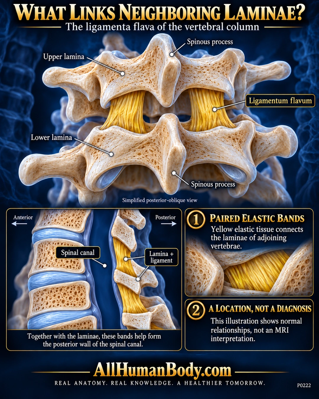
The ligamenta flava are paired elastic-tissue bands connecting neighboring vertebral laminae. Together with these bony arches they contribute to the posterior wall of the spinal canal.
READ THE IMAGE
What the illustration shows
English anatomy poster on a navy field. An oblique posterior illustration shows two ivory vertebral arches connected by paired yellow bands between their laminae. Labels distinguish upper and lower laminae, ligamentum flavum and spinous processes. A sagittal inset places vertebral bodies and discs anteriorly, an empty blue spinal canal centrally, and laminae with short yellow ligaments posteriorly. Text describes paired elastic bands and states that the illustration is not an MRI interpretation. Gold AllHumanBody.com branding and full slogan form the footer.
How to interpret this poster
- Bone texture, ligament thickness and spacing are schematic, not a named vertebral level, exact attachment subdivision or surgical atlas.
- Elastic character does not quantify recoil, tissue percentage or motion range.
- No MRI interpretation, stenosis diagnosis, buckling demonstration or treatment advice.
- AI-assisted educational illustration; not physician certification, microscopy, a diagnosis or individualized medical advice.
- Fine annotations need enlargement; the full description provides an accessible text alternative.
- Display the full uncropped illustration; do not trim titles, legends or boundary annotations.
AI-assisted educational illustration, adapted into web-sized images without changing its composition. This is general education, not an individual diagnosis or treatment recommendation.
EVIDENCE / CONTEXT
Sources and what they support
- NLM MeSH: Ligamentum Flavum ↗
Paired elastic bands joining adjacent laminae and contributing to posterior canal wall.