Musculoskeletal / VISUAL EXPLAINER
Where Do Spinal Nerves Leave the Bony Spine?
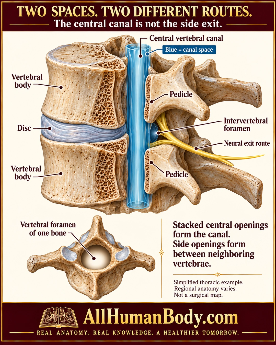
Stacked vertebral openings form a central canal, while neighboring vertebrae form separate side openings called intervertebral foramina. Neural structures use these lateral passages; a highlighted canal space is not itself a nerve or blood vessel.
READ THE IMAGE
What the illustration shows
Portrait dimensional cutaway of two neighboring vertebrae with a grey disc between their bodies. A blue highlighted vertical space behind the bodies is labeled central vertebral canal and explicitly identified as canal space. One yellow neural route leaves through the side opening between neighboring pedicles, labeled intervertebral foramen. A lower inset shows the central vertebral foramen of a single bone from above. Text distinguishes stacked central openings from side openings and notes regional variation. Gold AllHumanBody.com and full slogan are intact.
How to interpret this poster
- Simplified thoracic example, not exact regional root anatomy, meninges or a surgical section.
- Blue is a space marker; dimensions and exposed pedicle shapes are illustrative.
- No compression diagnosis, injection path, universal nerve numbering or operative guidance.
- AI-assisted educational illustration; not physician certification, microscopy, a diagnosis or individualized medical advice.
- Fine annotations need enlargement; the full description provides an accessible text alternative.
- Display the full uncropped illustration; do not trim titles, legends or boundary annotations.
AI-assisted educational illustration, adapted into web-sized images without changing its composition. This is general education, not an individual diagnosis or treatment recommendation.
EVIDENCE / CONTEXT
Sources and what they support
- Anatomy and Physiology 2e: 7.3 The Vertebral Column ↗
Central vertebral openings and lateral intervertebral passages; directly read general structure section preserved and reopened during production.