Digestive / VISUAL EXPLAINER
Which Cells Make the Mucus on Your Intestinal Lining?
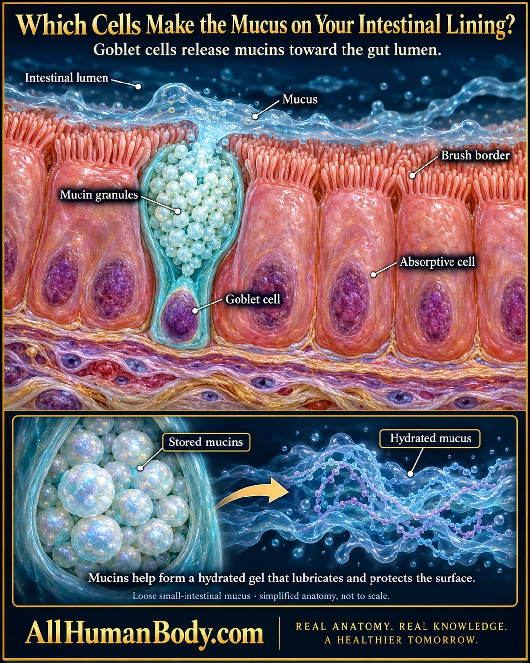
Goblet cells contribute mucins to the intestinal surface. Their secretion helps form mucus that lubricates and supports the mucosal barrier, alongside the other roles of neighboring epithelial cells.
READ THE IMAGE
What the illustration shows
Original educational poster showing an enlarged small-intestinal epithelial surface. A teal goblet cell sits between pink absorptive cells with short brush-border microvilli. Its broad upper cup contains pale mucin granules, and its narrow base contains a purple nucleus. Translucent blue mucus leaves the cell toward the lumen and spreads over the surface. A lower inset contrasts stored granules with a hydrated, loosely drawn polymer gel. The gel is an artistic teaching representation, not a molecular model. Text explains lubrication and protection, with a simplified-anatomy caveat. Gold AllHumanBody.com branding and the full project slogan appear below.
How to interpret this poster
- The blue mesh is a mucus-gel symbol, not a molecular structure or impermeable wall.
- Short brush borders are microvilli, not beating cilia.
- No fixed human turnover rate, colon-like two-layer claim or mucus treatment.
- AI-assisted educational illustration; not physician certification, microscopy, a diagnosis or individualized medical advice.
- Fine annotations need enlargement; the full description provides an accessible text alternative.
- Display the full uncropped illustration; do not trim titles, legends or boundary annotations.
AI-assisted educational illustration, adapted into web-sized images without changing its composition. This is general education, not an individual diagnosis or treatment recommendation.
EVIDENCE / CONTEXT
Sources and what they support
- University of Leeds Histology Guide: Small Intestine ↗
Goblet-cell mucus and neighboring absorptive lining.
- Study of mucin turnover in the small intestine by in vivo labeling ↗
Stored mucin secretion and small-intestinal mucus context in mice.
Evidence correspondence is limited to the abstract.