Digestive / VISUAL EXPLAINER
What Anchors Your Small-Intestinal Loops?
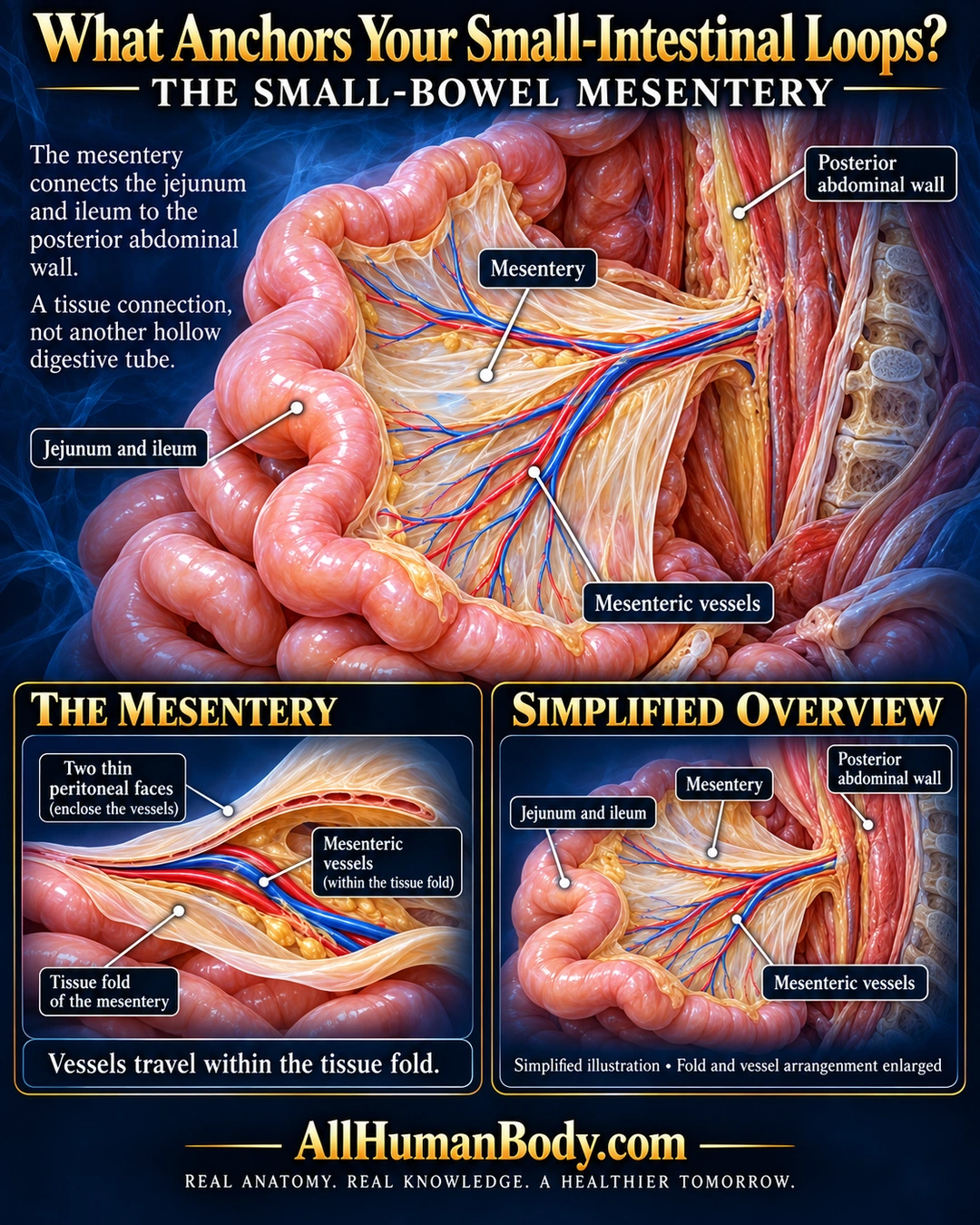
The small-bowel mesentery connects the jejunum and ileum to the posterior abdominal wall. Blood vessels travel within this supporting tissue fold, rather than suspending the bowel as bare vessels or forming another digestive tube.
READ THE IMAGE
What the illustration shows
English medical poster on navy background. An oblique abdominal view shows pink small-bowel loops attached by a fan-shaped ivory mesenteric tissue fold to the posterior abdominal wall at right. Red and blue vessels branch within the fold. Labels identify jejunum and ileum, mesentery, vessels and posterior wall. Lower left enlarged section separates two peritoneal faces to reveal enclosed vessels; lower right repeats a simplified overview. Tissue spacing and vessel arrangement are enlarged, not exact branching or a fixed loop configuration. Gold AllHumanBody.com and full slogan below.
How to interpret this poster
- Fold spacing and branching are enlarged, not a fixed bowel configuration or exact vascular tree. No guarantee against twisting or obstruction and no treatment advice.
- AI-assisted educational illustration; not physician certification, microscopy, a diagnosis or individualized medical advice.
- Fine annotations need enlargement; the full description provides an accessible text alternative.
- Display the full uncropped illustration; do not trim titles, legends or boundary annotations.
AI-assisted educational illustration, adapted into web-sized images without changing its composition. This is general education, not an individual diagnosis or treatment recommendation.
EVIDENCE / CONTEXT
Sources and what they support
- Texas Tech medical anatomy tables ↗
Mesentery attachment and intestinal arteries within it.
- Texas Tech anatomical dissection teaching ↗
Small-bowel mesentery body-wall attachment and vessels.