Digestive / VISUAL EXPLAINER
What Makes the Duodenum Different Under the Microscope?
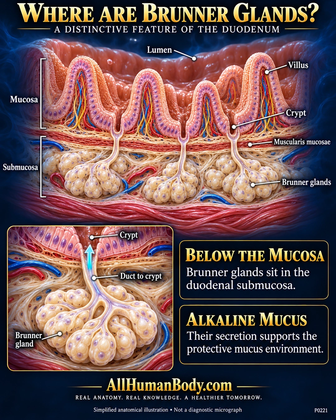
Brunner glands sit in the duodenal submucosa beneath the muscularis mucosae. Their ducts reach the crypts above, providing a route for alkaline mucus to enter the intestinal lumen.
READ THE IMAGE
What the illustration shows
English anatomy poster on a navy field. Peach duodenal villi rise above crypts and a thin red muscularis mucosae. Ivory Brunner gland lobules occupy the submucosa below; narrow pale ducts connect them to crypts. A lower-left enlargement follows a duct upward with a cyan arrow. Two text panels identify submucosal location and alkaline mucus. Gold AllHumanBody.com branding and full slogan appear at the bottom. Simplified illustration, not a diagnostic micrograph.
How to interpret this poster
- Lobule counts, cell shapes, vessel patterns and layer thickness are schematic, not a diagnostic micrograph.
- No exact pH, guaranteed acid protection, treatment or claim that these glands produce pancreatic bicarbonate.
- No absolute distribution boundary that excludes all anatomical variation.
- AI-assisted educational illustration; not physician certification, microscopy, a diagnosis or individualized medical advice.
- Fine annotations need enlargement; the full description provides an accessible text alternative.
- Display the full uncropped illustration; do not trim titles, legends or boundary annotations.
AI-assisted educational illustration, adapted into web-sized images without changing its composition. This is general education, not an individual diagnosis or treatment recommendation.
EVIDENCE / CONTEXT
Sources and what they support
- Histology Guide: MHS272 Duodenum ↗
Submucosal location, muscularis mucosae boundary and alkaline mucus.
- Loyola University HistoLab: Part17, Digestive System ↗
Brunner ducts cross muscularis mucosae into crypts, continuous with intestinal lumen.