Digestive / VISUAL EXPLAINER
Is the Space Between Intestinal Cells Completely Sealed?
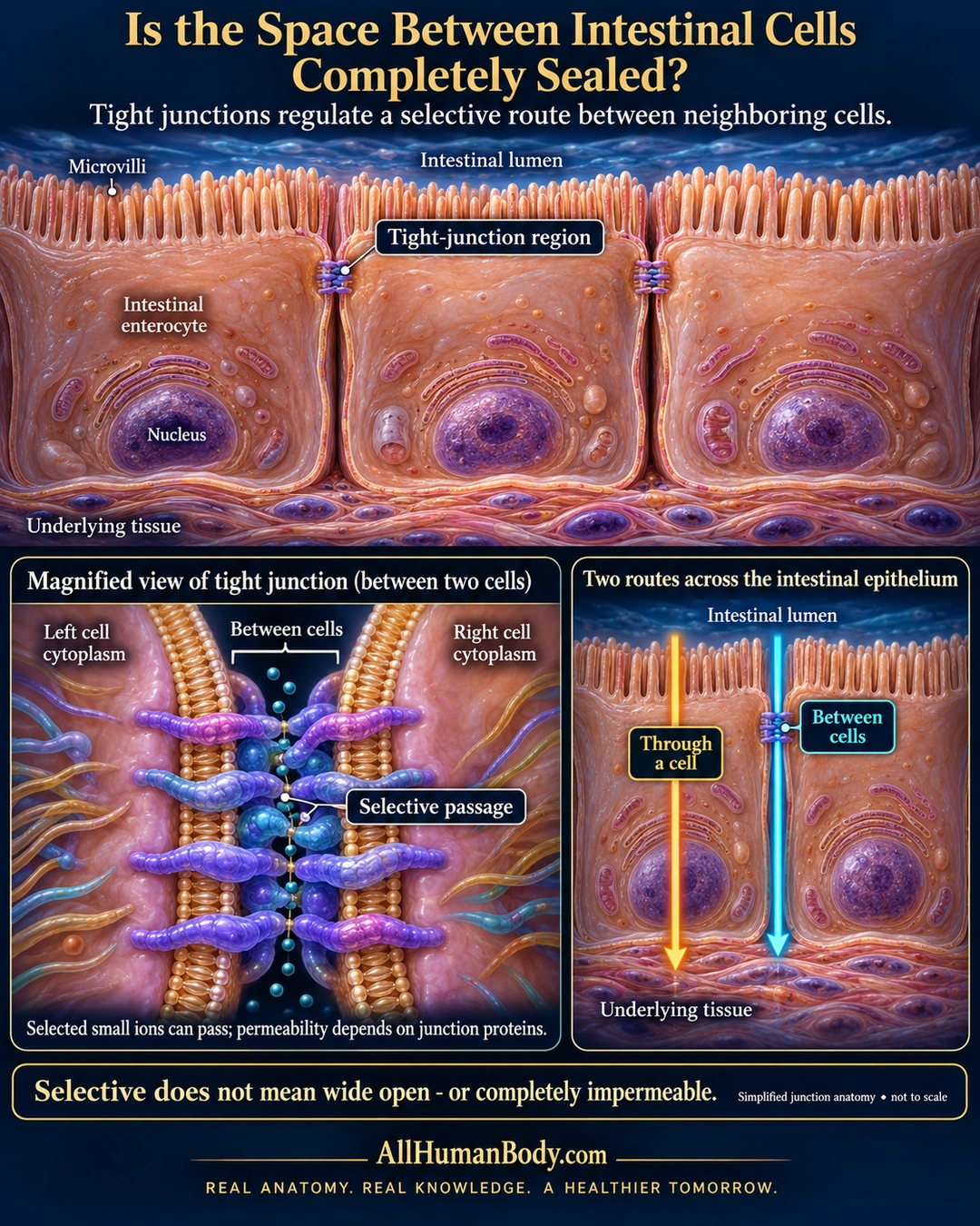
Tight junctions near the apical edges of intestinal epithelial cells regulate passage between neighboring cells. This paracellular route is selective, rather than a permanently open gap or a completely impermeable wall.
READ THE IMAGE
What the illustration shows
English medical poster about selective passage between intestinal cells. Three dimensional enterocytes have microvilli facing the intestinal lumen, basal nuclei and highlighted tight-junction regions near their upper lateral edges. Beneath them is underlying tissue. A magnified inset shows opposing membranes, stylized protein contacts and tiny ion dots along a narrow intercellular route, labeled selective passage. A second inset contrasts a gold arrow through one cell with a turquoise arrow between adjacent cells. Text says permeability depends on junction proteins and selective does not mean wide open or completely impermeable. Anatomy and molecular contacts are schematic, not to scale. Gold AllHumanBody.com and its full slogan appear at the bottom.
How to interpret this poster
- Protein contacts and ion spacing are symbols, not measured pores or molecular structure.
- No universal pore size, absolute water blockade or claim that bacteria freely cross normal epithelium.
- No leaky-gut diagnosis, supplement-repair or treatment claim.
- AI-assisted educational illustration; not physician certification, microscopy, a diagnosis or individualized medical advice.
- Fine annotations need enlargement; the full description provides an accessible text alternative.
- Display the full uncropped illustration; do not trim titles, legends or boundary annotations.
AI-assisted educational illustration, adapted into web-sized images without changing its composition. This is general education, not an individual diagnosis or treatment recommendation.
EVIDENCE / CONTEXT
Sources and what they support
- Claudins and the modulation of tight junction permeability ↗
Apical junctional organization and selective paracellular barriers and pores.