Digestive / VISUAL EXPLAINER
Which Cells Release Defenses at the Bottom of Intestinal Crypts?
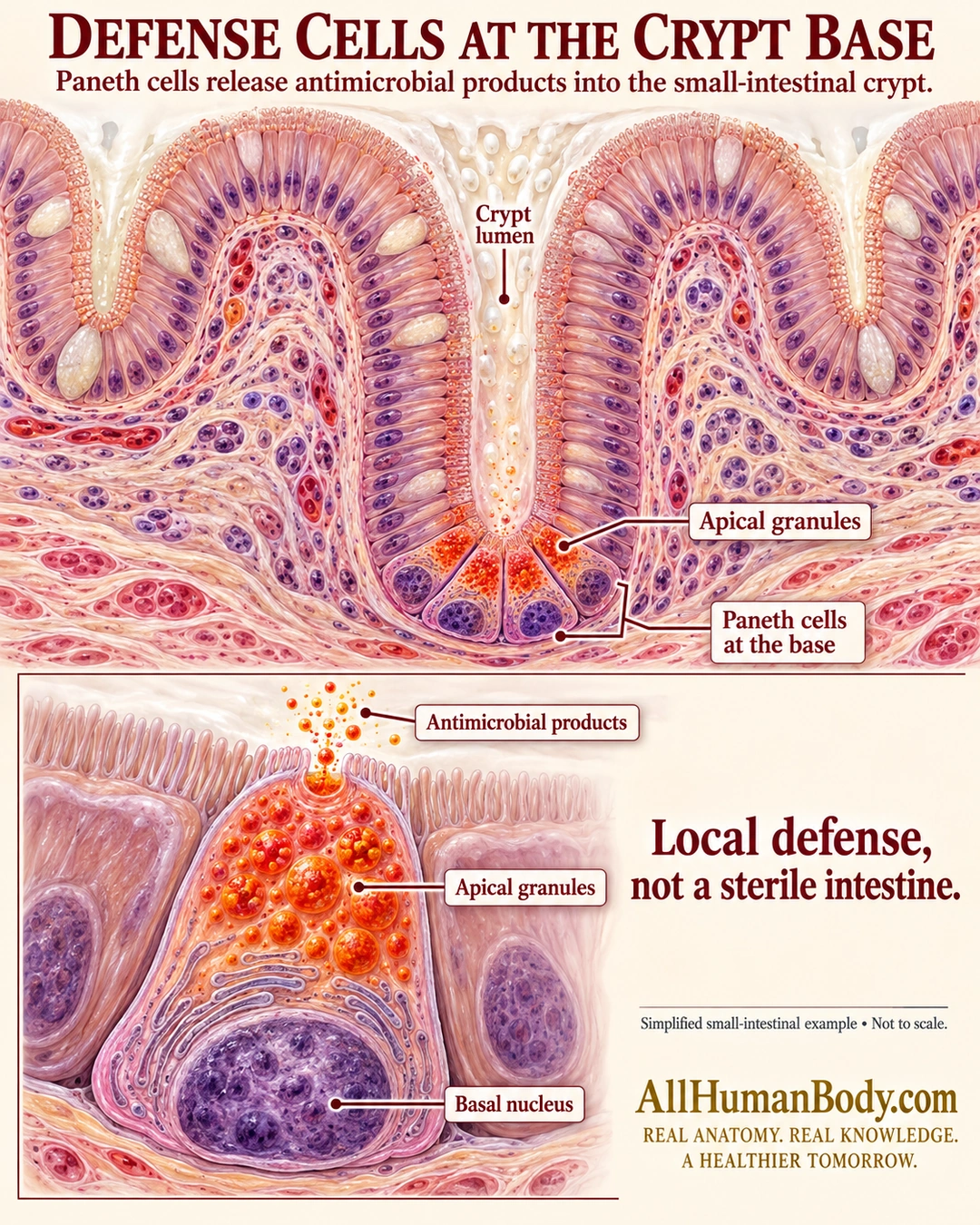
Paneth cells at the base of small-intestinal crypts contain apical granules with antimicrobial products. Release into the crypt lumen contributes to local defense without making the intestine sterile.
READ THE IMAGE
What the illustration shows
A dimensional tissue poster shows a small-intestinal crypt open upward between neighboring mucosal profiles. Paneth cells at its base have purple basal nuclei and orange-red granules concentrated toward the crypt lumen. A lower enlarged cell shows an apical fusion opening releasing small products outward into the lumen, with the nucleus at the opposite basal end. Labels identify the crypt lumen, Paneth cells, apical granules, antimicrobial products and basal nucleus. Text reads Local defense, not a sterile intestine. Gold AllHumanBody.com and its full slogan appear below.
How to interpret this poster
- Selected cell profiles and granule dots are schematic, not measured histology, defensin structures or cell counts.
- No normal-colon or villus-tip Paneth claim, fixed turnover time or disease diagnosis.
- No antimicrobial treatment, supplement or sterilization promise.
- AI-assisted educational illustration; not physician certification, microscopy, a diagnosis or individualized medical advice.
- Fine annotations need enlargement; the full description provides an accessible text alternative.
- Display the full uncropped illustration; do not trim titles, legends or boundary annotations.
AI-assisted educational illustration, adapted into web-sized images without changing its composition. This is general education, not an individual diagnosis or treatment recommendation.
EVIDENCE / CONTEXT
Sources and what they support
- University of Leeds Histology Guide: Small intestine ↗
Crypt-base Paneth cells and antimicrobial granule contents.