Digestive / VISUAL EXPLAINER
How Does the Gut Sample What Is Inside Its Lumen?
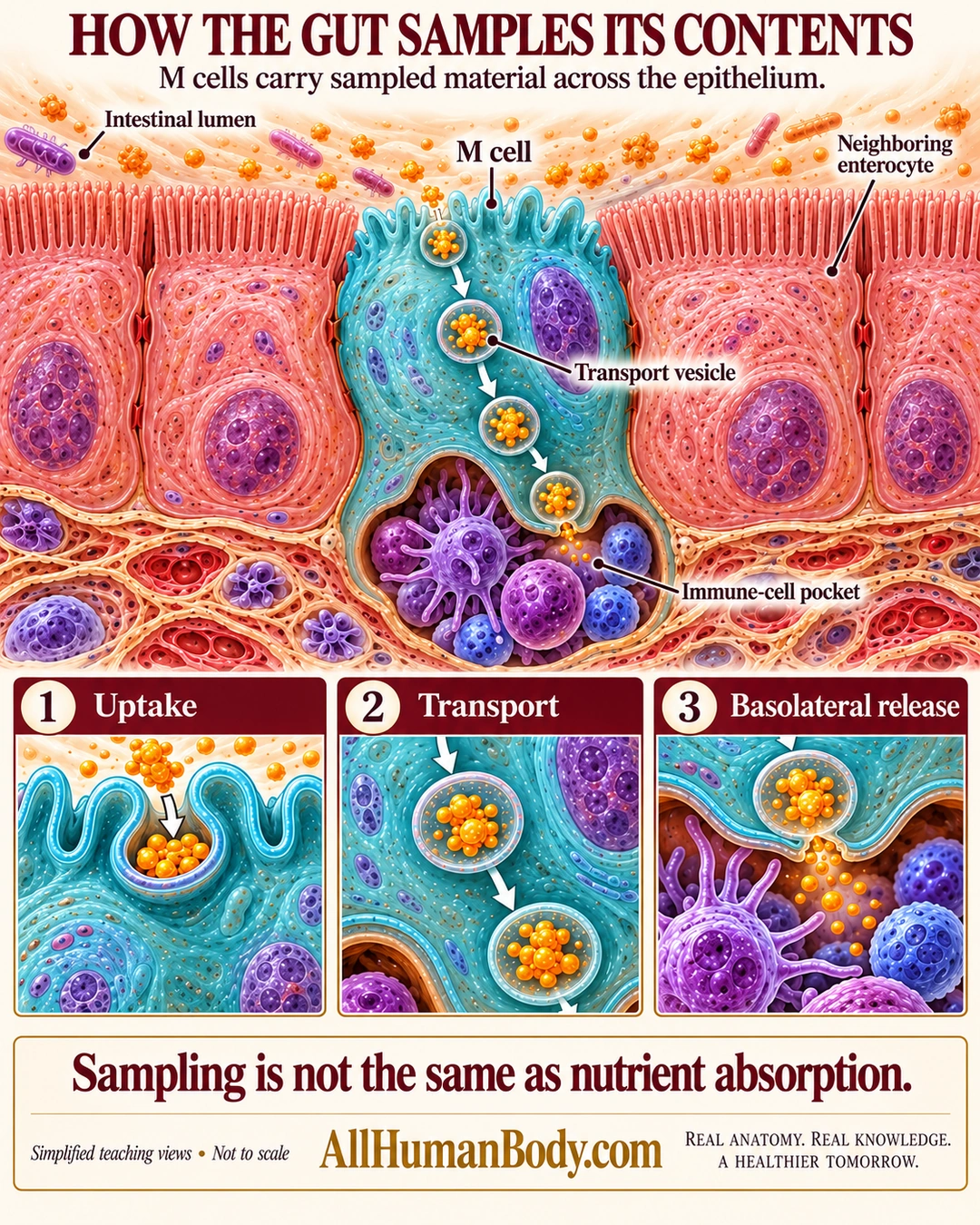
M cells in epithelium over intestinal lymphoid tissue can move sampled luminal material across the cell toward local immune cells. This epithelial sampling role differs from the main nutrient-absorbing role of neighboring enterocytes.
READ THE IMAGE
What the illustration shows
Cream medical teaching poster titled How the gut samples its contents. A teal M cell lies between pink enterocytes beneath the intestinal lumen. Orange sample particles are enclosed in transport vesicles within the M cell and released at its indented basal side toward purple immune cells in a pocket. The neighboring enterocytes have prominent brush borders. Three lower panels show uptake, transport and basolateral release. The takeaway distinguishes sampling from nutrient absorption. Tissue colors, shapes and proportions are schematic. Gold AllHumanBody.com branding and the full slogan appear at the bottom.
How to interpret this poster
- Qualitative teaching model, not direct human microscopy: cited morphology includes rat specimen and human organoid research.
- Surface folds, vesicle numbers, pocket cell mixture and proportions are schematic; surrounding lymphoid architecture is omitted.
- No open lumen-to-blood pipe, guaranteed sterilization, pathogens-only sampling, claim M cells cannot present antigen, or coeliac/vaccine/treatment inference.
- AI-assisted educational illustration; not physician certification, microscopy, a diagnosis or individualized medical advice.
- Fine annotations need enlargement; the full description provides an accessible text alternative.
- Display the full uncropped illustration; do not trim titles, legends or boundary annotations.
AI-assisted educational illustration, adapted into web-sized images without changing its composition. This is general education, not an individual diagnosis or treatment recommendation.
EVIDENCE / CONTEXT
Sources and what they support
- Histology Guide: EM 054 — M Cell ↗
Epithelial location and basal immune-cell pocket, comparative rat morphology.
- Human gut M cells resemble dendritic cells and present gluten antigen ↗
Human organoid antigen transport with broader M-cell background; demonstrates why inability to present antigen is not a safe absolute.