Digestive / VISUAL EXPLAINER
Does Your Intestinal Wall Have Its Own Nerve Networks?
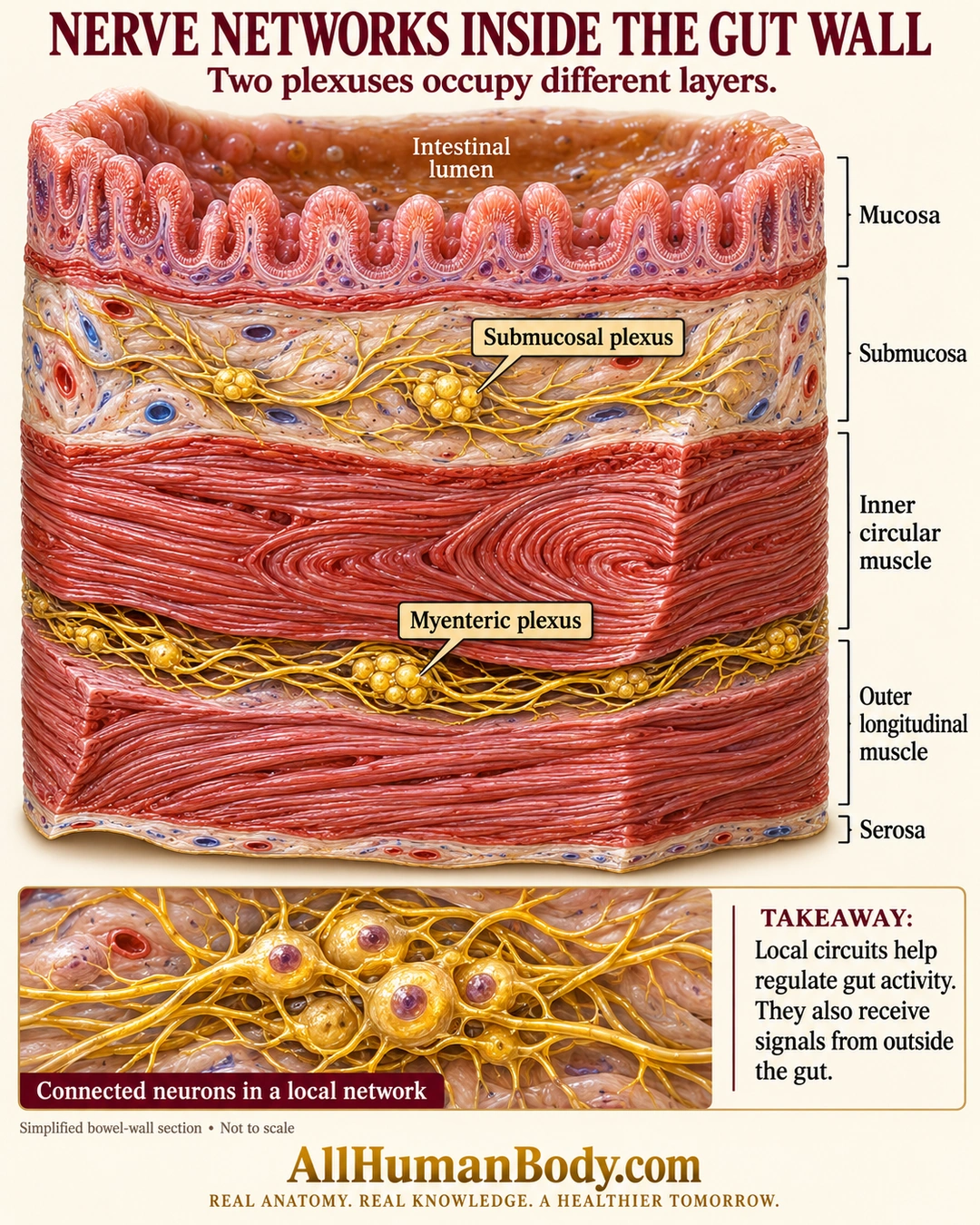
Enteric nerve networks lie within the bowel wall. The submucosal plexus occupies the submucosa, while the myenteric plexus lies between the main circular and longitudinal muscle layers; local regulation also interacts with signals from outside the gut.
READ THE IMAGE
What the illustration shows
Ivory medical poster shows a layered intestinal-wall cutaway with lumen and mucosa above loose submucosal connective tissue. A gold submucosal nerve network lies in that layer. Below it are inner circular muscle, a gold myenteric network between muscle layers, outer longitudinal muscle and serosa. A lower enlargement shows connected neuronal cell bodies and fibers. Layer labels and a takeaway describe local regulation and outside signals. Tissue proportions and fiber textures are simplified and not to scale. Gold AllHumanBody.com branding and full slogan complete the poster.
How to interpret this poster
- Ganglia enlarged and muscle textures stylized; not exact fiber directions, thicknesses or a complete reflex circuit.
- Thin muscularis mucosae is visible but not separately labeled.
- No second thinking brain, neuron count, microbiome-behavior claim or treatment advice.
- AI-assisted educational illustration; not physician certification, microscopy, a diagnosis or individualized medical advice.
- Fine annotations need enlargement; the full description provides an accessible text alternative.
- Display the full uncropped illustration; do not trim titles, legends or boundary annotations.
AI-assisted educational illustration, adapted into web-sized images without changing its composition. This is general education, not an individual diagnosis or treatment recommendation.
EVIDENCE / CONTEXT
Sources and what they support
- Anatomy and Physiology 2e: 23.1 Overview of the Digestive System ↗
Wall layers, intrinsic plexuses, local regulation and extrinsic nerve input; preserved direct Nerve Supply and layer sections.
- Southern Illinois University Histology: Myenteric plexus ↗
Myenteric plexus between circular and longitudinal muscle.