Musculoskeletal / VISUAL EXPLAINER
What Holds Your Hyoid Bone in Place?
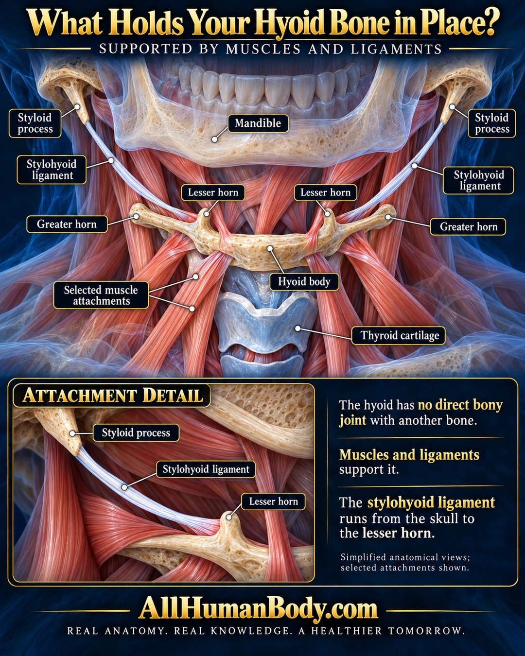
The hyoid lies below the jaw and above the thyroid cartilage. Muscles and ligaments support it without a direct bony joint; the stylohyoid ligament links the skull’s styloid process to the hyoid’s lesser horn.
READ THE IMAGE
What the illustration shows
English anatomy poster titled “What Holds Your Hyoid Bone in Place?” A frontal neck cutaway shows a translucent mandible above an ivory hyoid and blue-gray thyroid cartilage below. Paired white stylohyoid ligaments descend from skull styloid processes toward the small lesser horns; larger horns extend outward and backward in the frontal projection. Peach-red muscle slips surround and attach above and below the hyoid. Leaders label the mandible, styloid processes, ligaments, lesser and greater horns, hyoid body, selected muscles and thyroid cartilage. A lower enlarged detail follows one ligament from its styloid process to the lesser horn. Text explains soft-tissue support without a direct bony joint. Navy and cyan setting, gold brand and full slogan.
How to interpret this poster
- Selected supports only, not a freely floating bone or complete muscle map. No fixed vertebral level, forensic inference or manipulation guidance.
- AI-assisted educational illustration; not physician certification, microscopy, a diagnosis or individualized medical advice.
- Fine annotations need enlargement; the full description provides an accessible text alternative.
- Display the full uncropped illustration; do not trim titles, legends or boundary annotations.
AI-assisted educational illustration, adapted into web-sized images without changing its composition. This is general education, not an individual diagnosis or treatment recommendation.
EVIDENCE / CONTEXT
Sources and what they support
- Academic medical center patient anatomy ↗
Position, shape, and soft-tissue support. Vertebral-level and fusion-age claims excluded.
- StatPearls anatomy reference ↗
Styloid apex and lesser-cornu attachment of stylohyoid ligament.