Musculoskeletal / VISUAL EXPLAINER
Which Bones Form the Hard Palate?
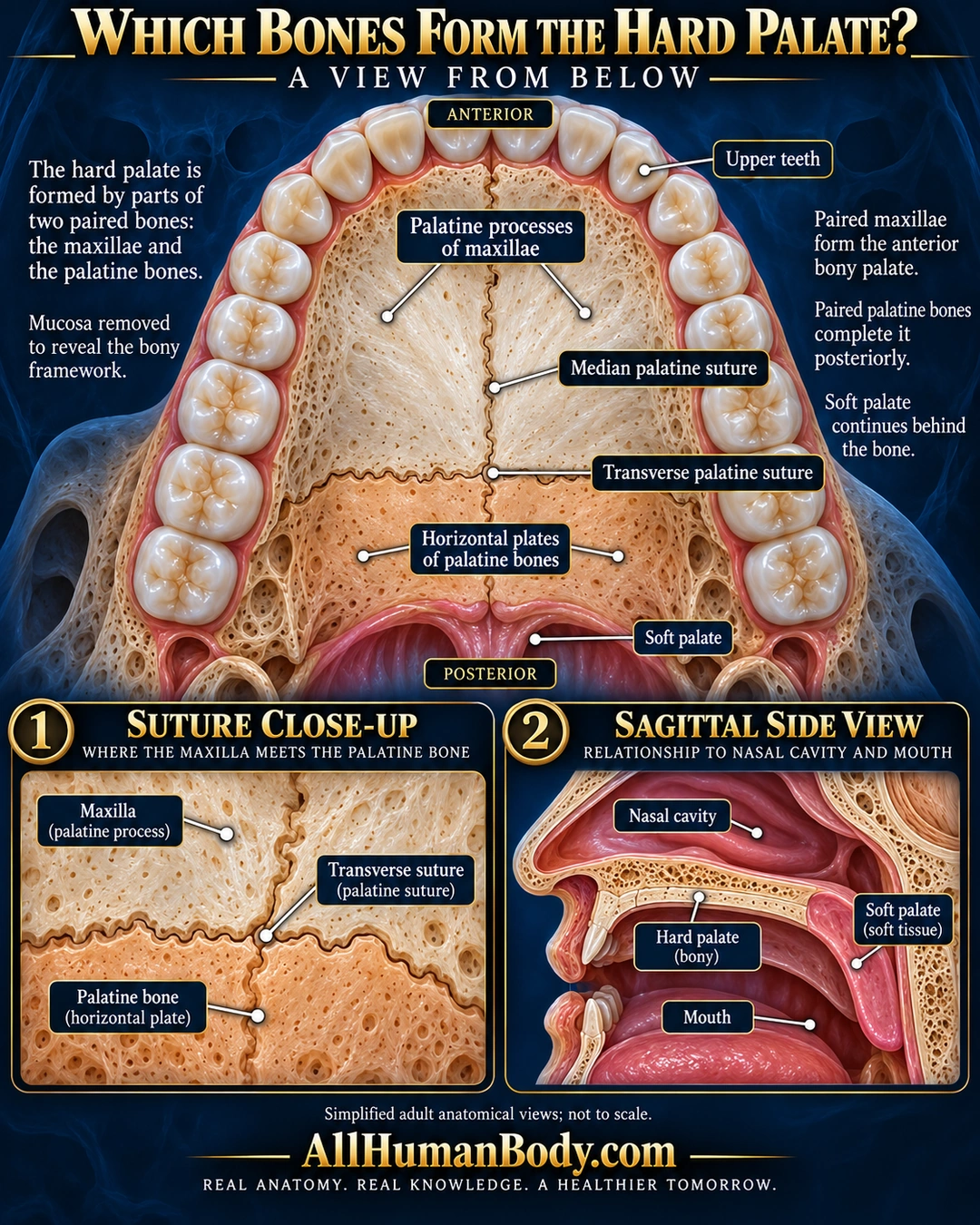
The hard palate has a bony framework made from paired maxillary palatine processes at the front and paired horizontal palatine plates farther back. The soft palate continues behind this framework as soft tissue.
READ THE IMAGE
What the illustration shows
English anatomy poster titled “Which Bones Form the Hard Palate?” An inferior upper-jaw view places the front teeth at the top. Ivory paired maxillary palatine processes occupy the anterior palate and peach-tinted paired horizontal palatine plates occupy the posterior part. Midline and transverse sutures separate the regions. Paired leaders point to both halves, and a pink soft-palate continuation sits behind the bony edge. Labels identify upper teeth and orientation. Below, a suture close-up identifies maxilla and palatine bone; a sagittal locator places the hard palate between nasal cavity and mouth and shows soft tissue continuing posteriorly. Navy-blue background, gold brand and full slogan.
How to interpret this poster
- Mucosa removed in teaching view; generated sutures and regional proportions are illustrative adult anatomy. No cleft-development, growth or treatment claims.
- AI-assisted educational illustration; not physician certification, microscopy, a diagnosis or individualized medical advice.
- Fine annotations need enlargement; the full description provides an accessible text alternative.
- Display the full uncropped illustration; do not trim titles, legends or boundary annotations.
AI-assisted educational illustration, adapted into web-sized images without changing its composition. This is general education, not an individual diagnosis or treatment recommendation.
EVIDENCE / CONTEXT
Sources and what they support
- NCI SEER anatomy teaching ↗
Oral/nasal separation and bony versus soft posterior palate.
- Cincinnati Children’s faculty teaching handout by Ann W. Kummer ↗
Maxillary palatine processes, palatine horizontal plates, transverse suture, posterior soft-palate attachment.