Respiratory / VISUAL EXPLAINER
Is Your Nasal Septum All Cartilage?
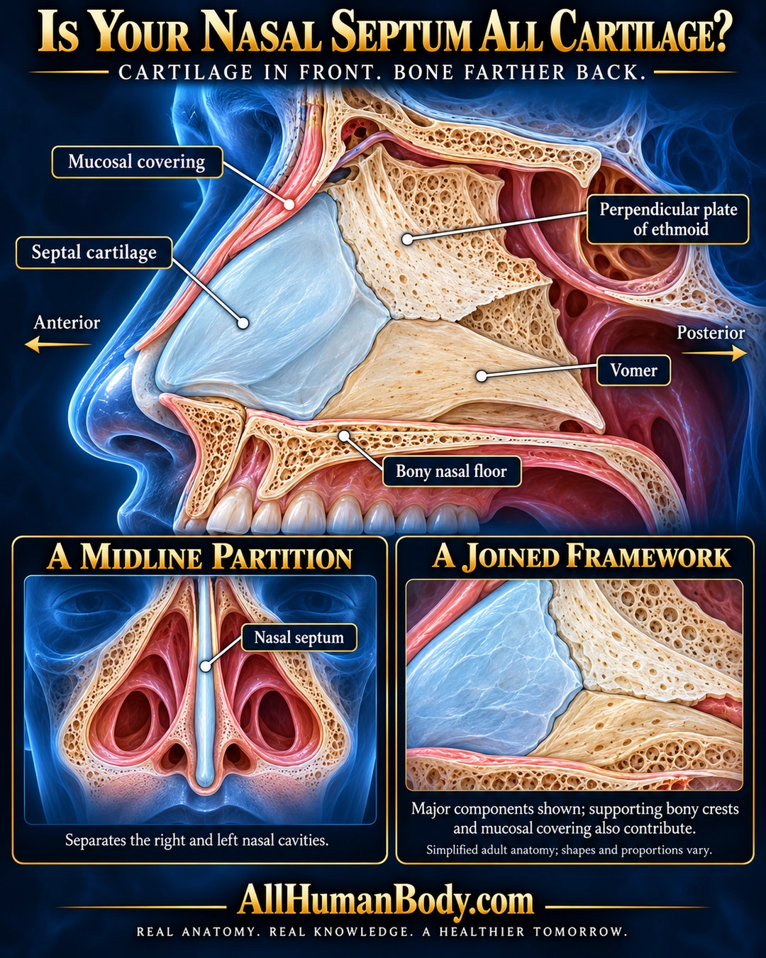
The nasal septum separates the two nasal cavities. Its front is supported by cartilage, while the ethmoid and vomer contribute major bony parts farther back; covering tissue and supporting bony crests also contribute.
READ THE IMAGE
What the illustration shows
English anatomy poster titled “Is Your Nasal Septum All Cartilage?” on a deep navy background. A large sagittal head view faces left. Pale cyan septal cartilage is anterior, with an ivory perpendicular plate of the ethmoid above and a warmer ivory vomer below and behind it. Leaders label these components, the bony nasal floor and a pink mucosal covering. Anterior and posterior arrows orient the view. A frontal inset locates the central nasal septum between the nasal cavities; a second inset enlarges the joined cartilage and bone. Text notes that supporting bony crests and mucosal covering also contribute and that adult shapes and proportions vary. Gold AllHumanBody.com branding and the full project slogan appear below.
How to interpret this poster
- Representative adult shapes and proportions, not a perfectly straight universal septum or a complete component inventory. No deviation diagnosis or manipulation instructions.
- AI-assisted educational illustration; not physician certification, microscopy, a diagnosis or individualized medical advice.
- Fine annotations need enlargement; the full description provides an accessible text alternative.
- Display the full uncropped illustration; do not trim titles, legends or boundary annotations.
AI-assisted educational illustration, adapted into web-sized images without changing its composition. This is general education, not an individual diagnosis or treatment recommendation.
EVIDENCE / CONTEXT
Sources and what they support
- Stanford Medicine Sinus Center patient anatomy guide ↗
Midline partition, anterior cartilage, posterior bone, named contributors.