Special Senses / VISUAL EXPLAINER
How Does a Smell Become a Nerve Signal?
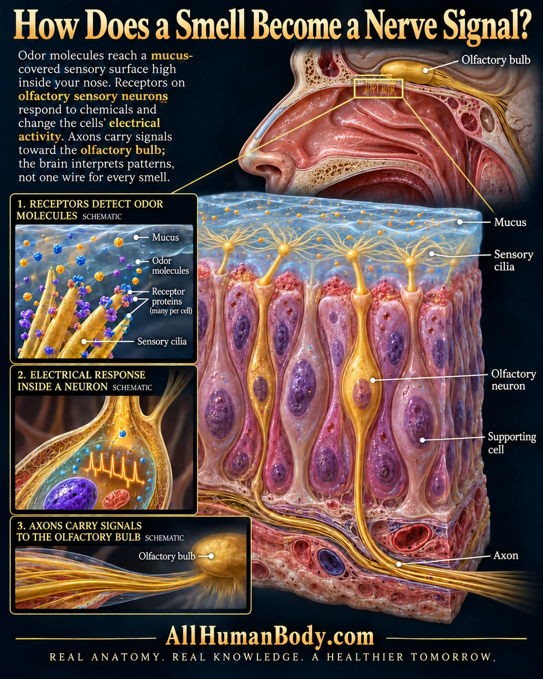
Odor molecules reach a mucus-covered sensory surface high inside the nose. Receptor activation changes olfactory-neuron electrical activity, and axons carry signals toward the bulb; smell depends on patterns of activity, not a separate wire for each odor.
READ THE IMAGE
What the illustration shows
A sagittal nose and brain locator highlights the olfactory region high in the nasal cavity. Below, a large tissue cutaway shows gold sensory neurons among pink supporting cells. Each highlighted neuron extends an apical process to a knob with fine sensory cilia spreading in blue mucus, and a basal axon joins a nerve-fiber bundle. Three schematic insets show odor molecules near receptor-bearing cilia, an illustrative electrical-activity symbol within a neuron, and fibers reaching the olfactory bulb. Text explains that the brain interprets patterns of signals, not a separate wire for every smell. Gold AllHumanBody.com branding appears below.
How to interpret this poster
- The waveform inside a neuron is an explanatory symbol, not a physical organelle or recording.
- No precise receptor map, glomerular wiring, cell count or guaranteed regeneration.
- Odor molecules are not shown traveling along axons into the brain; no smell-loss diagnosis.
- AI-assisted educational illustration; not physician certification, microscopy, a diagnosis or individualized medical advice.
- Fine annotations need enlargement; the full description provides an accessible text alternative.
- Display the full uncropped illustration; do not trim titles, legends or boundary annotations.
AI-assisted educational illustration, adapted into web-sized images without changing its composition. This is general education, not an individual diagnosis or treatment recommendation.
EVIDENCE / CONTEXT
Sources and what they support
- Anatomy and Physiology 2e: 14.1 Sensory Perception ↗
Bipolar olfactory neurons, mucus-covered apical processes and basal axons toward the bulb.
- Journey of Smell to the Brain: Text Version ↗
High nasal sensory neurons, patterned signals and brain interpretation.