Digestive / VISUAL EXPLAINER
Where Does a Pancreatic Duct Begin?
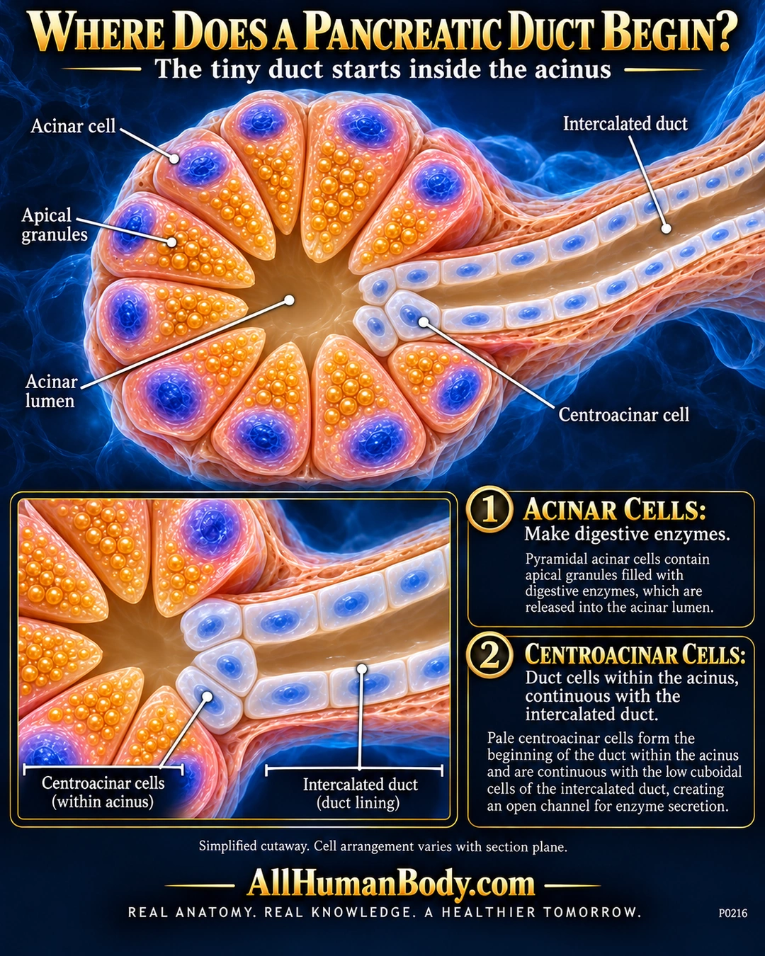
The pancreatic duct system begins within an acinus. Pale centroacinar cells are duct cells continuous with the intercalated duct, distinct from the surrounding acinar cells that make digestive secretory proteins.
READ THE IMAGE
What the illustration shows
Navy and gold pancreatic cutaway. Peach pyramidal acinar cells with blue basal nuclei and gold apical granules surround an open lumen. A small pale centroacinar cluster at the right joins the intercalated duct lining. Acinar apices directly face the lumen. A lower closeup repeats the transition; two text panels distinguish enzyme-producing acinar cells from duct cells. Gold AllHumanBody.com and its complete slogan appear on a plain navy footer.
How to interpret this poster
- Cell count, regular radial arrangement and lumen size are exaggerated; section-plane appearance varies and this is not a literal stained tissue field.
- Some stored digestive proteins are inactive precursors, not all active enzymes.
- Centroacinar cells are not endocrine islet cells or enzyme-making acinar cells; no treatment claim.
- AI-assisted educational illustration; not physician certification, microscopy, a diagnosis or individualized medical advice.
- Fine annotations need enlargement; the full description provides an accessible text alternative.
- Display the full uncropped illustration; do not trim titles, legends or boundary annotations.
AI-assisted educational illustration, adapted into web-sized images without changing its composition. This is general education, not an individual diagnosis or treatment recommendation.
EVIDENCE / CONTEXT
Sources and what they support
- Histology Guide: MH131 Pancreas ↗
Human pancreas description: duct origin within acinus, centroacinar identity and intercalated continuity.