Digestive / VISUAL EXPLAINER
Where Do Pancreatic Protein-Digesting Enzymes Switch On?
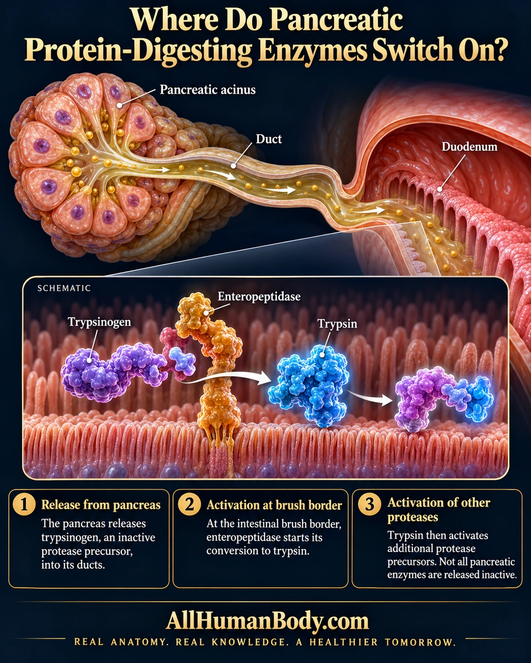
The pancreas releases trypsinogen through its exocrine ducts. At the intestinal brush border, enteropeptidase starts its conversion to trypsin, which can activate more protease precursors; not all pancreatic enzymes begin inactive.
READ THE IMAGE
What the illustration shows
Dark navy and gold anatomy poster asking where pancreatic protein-digesting enzymes switch on. An enlarged pancreatic acinus releases material through a duct toward a cutaway duodenum. Below, a labeled schematic shows purple trypsinogen beside surface-anchored enteropeptidase, a white arrow toward blue trypsin, and a further molecular interaction. Three text panels explain inactive precursor release, activation at the intestinal brush border, and activation of additional protease precursors. The poster notes that not all pancreatic enzymes are released inactive. AllHumanBody.com branding appears below.
How to interpret this poster
- The acinus-to-intestine picture combines microscopic and organ scales, not actual papilla geometry.
- The final molecular cluster represents an interaction, not an identified precursor or measured cleavage site.
- No pancreatitis diagnosis or enzyme-supplement guidance.
- AI-assisted educational illustration; not physician certification, microscopy, a diagnosis or individualized medical advice.
- Fine annotations need enlargement; the full description provides an accessible text alternative.
- Display the full uncropped illustration; do not trim titles, legends or boundary annotations.
AI-assisted educational illustration, adapted into web-sized images without changing its composition. This is general education, not an individual diagnosis or treatment recommendation.
EVIDENCE / CONTEXT
Sources and what they support
- Anatomy and Physiology 2e: 23.6 Accessory Organs in Digestion: The Liver, Pancreas, and Gallbladder ↗
Pancreatic protease precursors, intestinal enteropeptidase activation and amylase/lipase exceptions.