Lymphatic & Immune / VISUAL EXPLAINER
Are Tonsil Crypts the Same as Lymph-Node Channels?
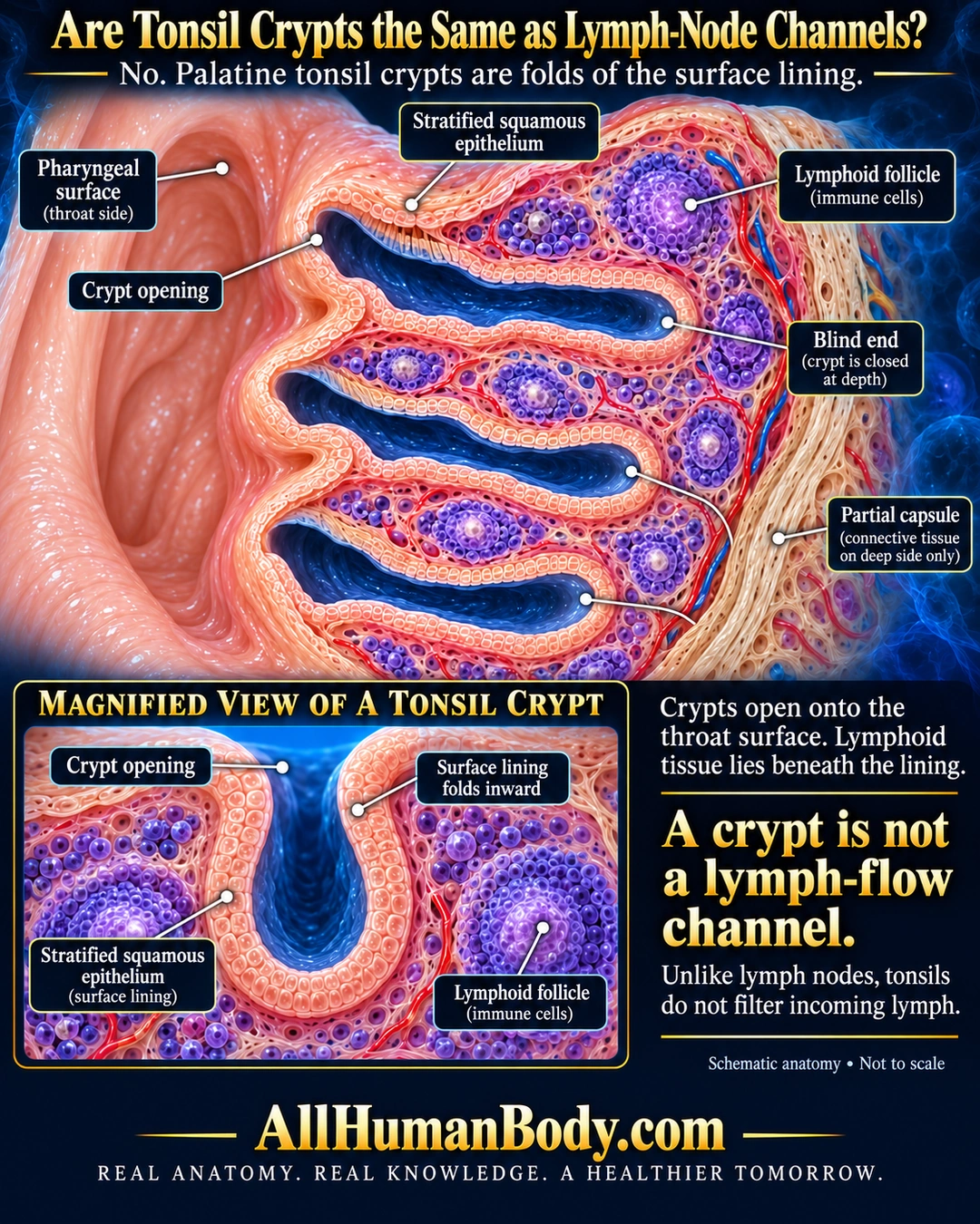
Palatine tonsil crypts are inward folds of surface epithelium, with lymphoid follicles beneath the lining. They connect to the throat surface and end blindly, unlike channels that bring incoming lymph through a lymph node.
READ THE IMAGE
What the illustration shows
English anatomy poster on a midnight-blue field. A palatine tonsil cutaway shows three blue crypt recesses opening left toward the throat and ending blindly within peach tissue. Purple lymphoid follicles sit beneath the lining; an ivory partial capsule borders the deep right side. Labels identify the pharyngeal surface, crypt opening, stratified squamous epithelium, follicles, blind ends and capsule. A lower magnification inset shows a U-shaped epithelial fold open at the top, with follicles outside it. Text contrasts crypts with lymph-flow channels. Gold AllHumanBody.com and the full brand slogan sit on a plain navy footer.
How to interpret this poster
- Restricted to palatine tonsil; deep squamous-lined crypts are not generalized to adenoids.
- Crypt count, folds, epithelial thickness and cell spacing are schematic. The orderly lining omits complex real lymphocyte infiltration.
- Blue shading denotes an open recess, not lymph flow. No tonsil-stone removal, infection diagnosis or treatment.
- AI-assisted educational illustration; not physician certification, microscopy, a diagnosis or individualized medical advice.
- Fine annotations need enlargement; the full description provides an accessible text alternative.
- Display the full uncropped illustration; do not trim titles, legends or boundary annotations.
AI-assisted educational illustration, adapted into web-sized images without changing its composition. This is general education, not an individual diagnosis or treatment recommendation.
EVIDENCE / CONTEXT
Sources and what they support
- University of Leeds Histology: Tonsils ↗
Blind epithelial crypts, underlying follicles and distinction from incoming-lymph filtration.
- Digital Histology: Palatine tonsil ↗
Palatine squamous epithelial invagination, lymphoid nodules and partial capsule.