Lymphatic & Immune / VISUAL EXPLAINER
How Do Antibodies Mark a Target?
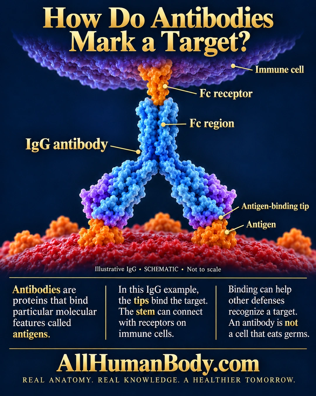
Antibodies bind particular molecular features called antigens. In this IgG example, binding tips recognize a target and the Fc region can interact with immune-cell receptors, helping coordinate other defenses rather than eating the target itself.
READ THE IMAGE
What the illustration shows
English poster titled How Do Antibodies Mark a Target? A large blue and purple IgG antibody forms an inverted Y. Its two lower antigen-binding tips contact orange features on a red target surface. Its upper Fc stem contacts an orange Fc receptor attached to a purple immune cell above. Leaders identify the immune cell, Fc receptor, Fc region, IgG antibody, antigen-binding tip and antigen. The diagram is marked illustrative, schematic and not to scale. Three text blocks explain recognition and cooperation with immune cells, noting that antibodies do not eat germs. Gold AllHumanBody.com and the project slogan appear below.
How to interpret this poster
- One illustrative IgG arrangement, not every antibody class or receptor interaction.
- Not atomic-scale geometry, a guaranteed clearance outcome or a complete immune pathway.
- The lesson covers recognition and cooperating defenses, not this full tip-and-stem molecular layout.
- AI-assisted educational illustration; not physician certification, microscopy, a diagnosis or individualized medical advice.
- Fine annotations need enlargement; the full description provides an accessible text alternative.
- Display the full uncropped illustration; do not trim titles, legends or boundary annotations.
AI-assisted educational illustration, adapted into web-sized images without changing its composition. This is general education, not an individual diagnosis or treatment recommendation.
EVIDENCE / CONTEXT
Sources and what they support
- Anatomy and Physiology 2e: 21.4 The Adaptive Immune Response: B-lymphocytes and Antibodies ↗
Antibody structure, antigen binding and Fc-region receptor interaction.