Digestive / VISUAL EXPLAINER
How Do Your Glands Make Saliva?
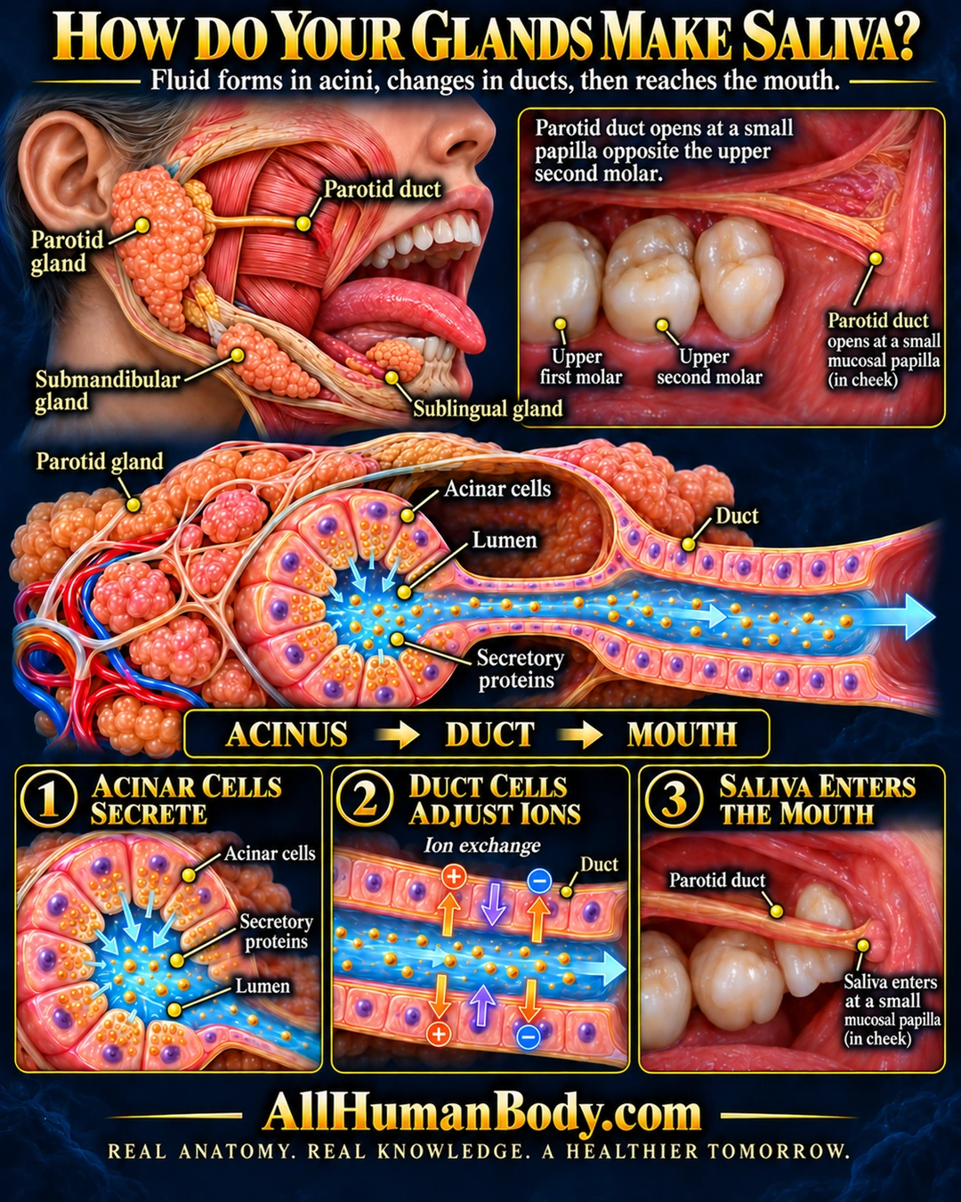
Saliva begins in secretory units called acini. In this parotid example, cells add fluid and proteins to the lumen, ducts modify the mixture, and a duct opening delivers it into the mouth.
READ THE IMAGE
What the illustration shows
Educational illustration titled “How Do Your Glands Make Saliva?” A head cutaway locates the parotid gland near the ear, submandibular gland below the jaw, and sublingual gland beneath the tongue. The highlighted parotid duct crosses cheek muscle and disappears beneath tissue. Mouth insets identify its small cheek papilla opposite the upper second molar. An enlarged parotid acinus shows cells releasing fluid and gold protein particles into a central lumen connected to a duct; blood vessels remain outside. Three panels summarize acinar secretion, duct ion exchange, and saliva entering the mouth. Blue arrows show saliva flow; separate charged symbols and mirrored arrows illustrate ion movement across duct walls. Footer: AllHumanBody.com.
How to interpret this poster
- A serous parotid example is shown, not the identical histology of every salivary gland.
- The hidden deep duct course and tooth-to-papilla relationship are schematic, not surgical localization.
- Ion arrows are qualitative and do not specify a full transporter map or fixed salivary composition.
- AI-assisted educational illustration; not physician certification, microscopy, a diagnosis or individualized medical advice.
- Fine annotations need enlargement; the full description provides an accessible text alternative.
AI-assisted educational illustration, adapted into web-sized images without changing its composition. This is general education, not an individual diagnosis or treatment recommendation.
EVIDENCE / CONTEXT
Sources and what they support
- Saliva & Salivary Gland Disorders ↗
Major-gland locations and delivery of saliva to the mouth.
- Anatomy and Physiology 2e: 23.3 The Mouth, Pharynx, and Esophagus ↗
Parotid secretion and its duct opening near the upper second molar.
- Salivary glands ↗
Secretory acini, protein-containing fluid and ductal modification of ions.