Digestive / VISUAL EXPLAINER
Which Cells Help a Salivary Gland Move Its Secretion?
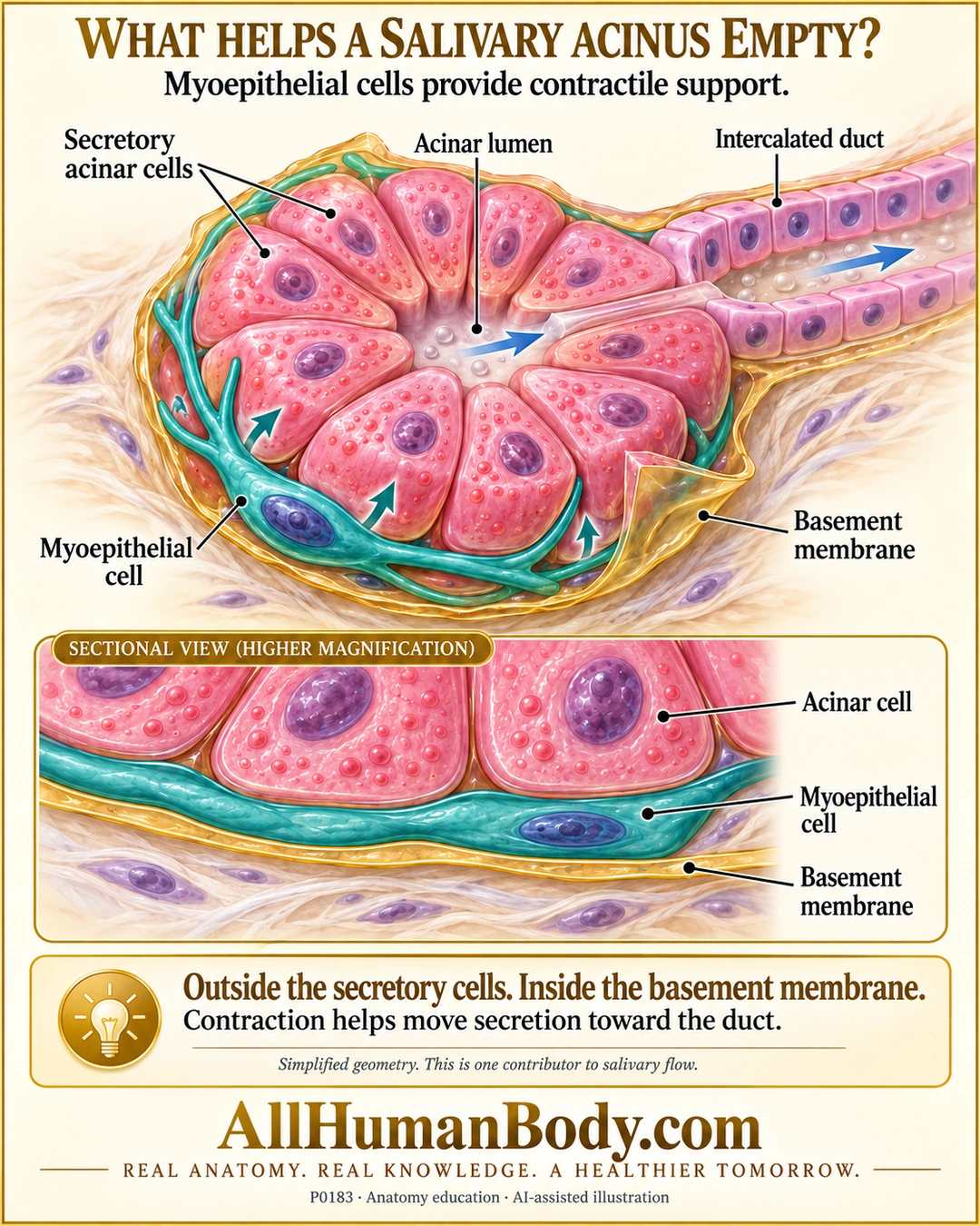
Salivary myoepithelial cells sit basal to secretory cells but inside their basement membrane. Their contractile processes help support the secretory unit and move secretion toward the duct, as one contributor to salivary flow.
READ THE IMAGE
What the illustration shows
Gold-and-ivory educational poster titled “What helps a salivary acinus empty?” A cutaway pink serous acinus surrounds a small central lumen opening into a cuboidal intercalated duct at upper right. Blue arrows follow the lumen toward the duct. A teal branching myoepithelial cell with a purple nucleus hugs the basal surface of the pink secretory cells, beneath a peeled transparent gold basement-membrane envelope. Teal arrows indicate contraction. A lower sectional inset repeats the order: pink acinar cells, teal myoepithelial cell, gold basement membrane, pale connective tissue. Text states that contraction helps secretion toward the duct and is one contributor to salivary flow. Gold AllHumanBody.com branding and the complete slogan appear below. Geometry, colors and layer thicknesses are illustrative, not measured anatomy.
How to interpret this poster
- One basket-like cell is not a measured cell count or complete wrapping pattern. Cell proportions, granules and basement-membrane thickness are illustrative.
- Arrows are not a quantitative force model, sole salivary pump or explanation of voluntary gleeking.
- The myoepithelial cell is not a nerve, skeletal-muscle bundle or the acinar cell producing secretion.
- AI-assisted educational illustration; not physician certification, microscopy, a diagnosis or individualized medical advice.
- Fine annotations need enlargement; the full description provides an accessible text alternative.
- Display the full uncropped illustration; do not trim titles, legends or boundary annotations.
AI-assisted educational illustration, adapted into web-sized images without changing its composition. This is general education, not an individual diagnosis or treatment recommendation.
EVIDENCE / CONTEXT
Sources and what they support
- University of Basel Histology: Parotid gland ↗
Basal myoepithelial location, acinar/intercalated-duct relationship and assistance in secretion expulsion.